Section Abstract Introduction Methods Results Discussion Conflict of Interest Acknowledgment References
Clinical Research
Genetic risk factor APOEε4 associates with plasma amyloid beta in amnestic mild cognitive impairment and alzheimer’s disease
pISSN: 0853-1773 • eISSN: 2252-8083
http://dx.doi.org/10.13181/mji.v25i1.1255 Med J Indones. 2016;25:44–50
Received: June 16, 2015
Accepted: February 08, 2016
Author affiliation:
1 Memory clinic Siloam Hospitals Lippo Village, Tangerang, Indonesia
2 Department of Neuroscience, Siloam Hospitals Lippo Village, Tangerang, Indonesia
3 Doctoral Program, Faculty of Medicine, Pelita Harapan University, Tangerang, Indonesia
4 Department of Neurology, Faculty of Medicine, Pelita Harapan University, Tangerang, Indonesia
5 Department of Neurology, Hasanuddin University, Makassar, Indonesia
6 Department of Pathology and Anatomy, Hasanuddin University, Makassar, Indonesia
7 Department of Neurology, Faculty of Medicine, Universitas Indonesia, Cipto Mangunkusumo Hospital, Jakarta, Indonesia
8 Doctoral Program, Hasanuddin University, Makassar, Indonesia
Corresponding author:
Rocksy F.V Situmeang
E-mail: rocksyfvs@yahoo.com
Background
APOEε4 is a strong genetic risk factor for Alzheimer’s disease (AD). AD itself has been associated with reduced Aβ clearance from the brain and plasma. Understanding the potential pathogenic link between APOEε4 and plasma Aβ might allow for earlier identification of people at risk of developing AD. The aim of this study is to find out the correlation between APOEε4 and plasma Aβ in amnestic mild cognitive impairment (aMCI) and AD patients.
Methods
This is a comparative cross-sectional study of patients attending a memory clinic in Siloam Hospital Lippo Karawaci, Tangerang, during the period of 2013-2014. Subjects were categorized into three categories: normal aging, aMCI, and AD. We performed blood test to examine APOEε4, plasma Aβ4o level, and plasma Aβ42 level. All data analyses were performed using correlation test and logistic regression.
Results
Sixty subjects (normal aging = 23, aMCI = 17, AD = 20) were included. There were 19 (31.7%) subjects with APOEε4 positive. Subjects carrying ε4 allele were more likely to have AD by 3.9-fold than subjects with APOE ε4 allele negative. There is a significant difference between the mean of plasma Aβ40 in aMCI group and AD group. We also found correlation between APOEε4 (+) and higher plasma Aβ42 (p<0.05).
Conclusion
There is a correlation between APOEε4 and plasma Aβ42 level, which supports the hypothesis that this genetic isoform accelerates the rate and progression of AD through Aβ-dependent pathways.
Keywords
alzheimer's disease, amnestic mild cognitive impairment, APOEε4, plasma amyloid beta
Alzheimer’s disease (AD) is the most common cause of dementia in the elderly. It is approximately occurred in 13% of people over the age of 65 to 45% people over the age of 85.1 One of the strongest pathomechanism of AD is amyloid cascade hypothesis that points to the imbalance between production and clearance of amyloid-β (Aβ) peptides in the brain which results in accumulation and aggregation of Aβ in the form of soluble Aβ oligomers, intraneuronal Aβ, and amyloid plaques injure synapses, and ultimately leads to neurodegeneration and dementia.2,3
The strongest risk factor for AD is the ε4 allele of the apolipoprotein E (APOE).4-6 There are three polymorphic alleles of APOE gene: ε2, ε3, and ε4, which APOEε3 allele is the most frequent in worldwide population (77.9%), followed by ε4 (13.7%), and ε2 (8.4%).7 Risk of AD was increased in individual with one copy of ε4 allele (heterozygote) and higher in two copies (homozygote).7 APOEε4 is associated with lower age of AD onset. Mean age at clinical AD onset in ε4 homozygote, ε4 heterozygote, and ε4 noncarriers are 68 years, 76 years, and 84 years, respectively.4,8
Mild cognitive impairment (MCI) is a transitional stage between normal cognitive function and dementia. Amnestic MCI (aMCI) is associated with increased risk of AD,9 is estimated to progress into clinically diagnosable AD in 10–15% of cases per year, compared to a rate of 1–2% per year among healthy elderly individuals.10 APOEε4 affects memory performance in people with MCI,11,12 both in middle age (40–59 years) and elderly (60–85 years) people.13,14 In addition, MCI patients with APOE ε4 experience more-rapid decline in several cognitive and functional assessments,12,15,16 which leads to increased risk of progression from MCI to AD-type dementia.17-19 Consistent with younger age of AD onset in individuals with APOEε4, it also applies in aMCI.20 These findings indicate that the APOEε4 genotype in patients with MCI can serve as a predictive factor for determination of clinical outcome and the risk of conversion to AD.21
APOE provides instruction to make a protein called Apo lipoprotein E- lipoproteins bind to several cell-surface receptors to deliver lipids and also to hydrophobic Aβ peptide, which is thought to initiate toxic events that lead to synaptic dysfunction and neurodegeneration in AD.21 APOE isoforms differentially regulate Aβ aggregation and clearance in the brain, and have distinct functions in regulating brain lipid transport, glucose metabolism, neuronal signalling, neuroinflammation, and mitochondrial function.21,22 Immunohistological evidence demonstrates that APOE is co-deposited in senile plaques in the brains of AD patients.23 The Aβ deposition in the form of senile plaques is more abundant in APOEε4 carriers compared with noncarriers.24-26 Furthermore, APOEε4 carriers have lower cerebrospinal fluid (CSF) Aβ42 level27,28 yet the relationship between plasma Aβ and APOEε4 remains inconclusive.29-32
In the present report, we studied the correlations between genetic risk factor APOEε4 with plasma amyloid beta level in both aMCI and AD.
METHODS
Subjects in this study were patients who visited memory clinic of Siloam Hospital Lippo Village from September 2013 to September 2014. We included all subjects who fulfilled inclusion criteria: 1) age >60 years old, 2) able to speak Bahasa Indonesia or English (premorbid), 3) able to read and write (premorbid). We excluded subjects with impaired renal function and subjects with major vascular lesions or other structural lesions in magnetic resonance imaging (MRI).
Subjects were categorized into three clinical diagnoses: normal aging, amnestic mild cognitive impairment, and AD. We performed physical, neurological, and neurobehavioral examinations including mini mental status examination (MMSE), montreal cognitive assessment (MoCA), and consortium to establish a registry of Alzheimer’s disease (CERAD). We used AD-8 questionnaire and instrumental activity daily living (IADL) to determine impairments on activities of daily living. We determined severity of AD using Clinical Dementia Rating Scale (CDRS) and Global deterioration Scale (GDS). MRI test was executed on subjects who had stroke history or suspected to have other causes of dementia.
We performed blood test to examine ureum, creatinine, APOEe4 with restriction fragment length polymorphism, plasma Aβ40 level, and plasma Aβ42 level. The concentration of Aβ peptides (Aβ40 and Aβ42) in plasma was measured using commercially available sandwich ELISA assay (IBL International GmbH, Hamburg, Germany) which has been shown to be sensitive enough 1.56–100 pg/mL.
All data analyses were performed using SPSS statistics (IBM Corp. Released 2011, version 20 Armonk, NY, US). The measures were expressed as mean ± standard deviation. We evaluated the association between two qualitative variables using chi square test and the association between qualitative variable and quantitative variable using t test. We performed the correlation test and logistic regression. A probability value less than 5% was considered statistically significant. The protocol of this study has been approved by Mochtar Riady Institute of Nanotechnology (No. 083/MRIN-EC/09/2014). We obtained written informed consent from all patients.
RESULTS
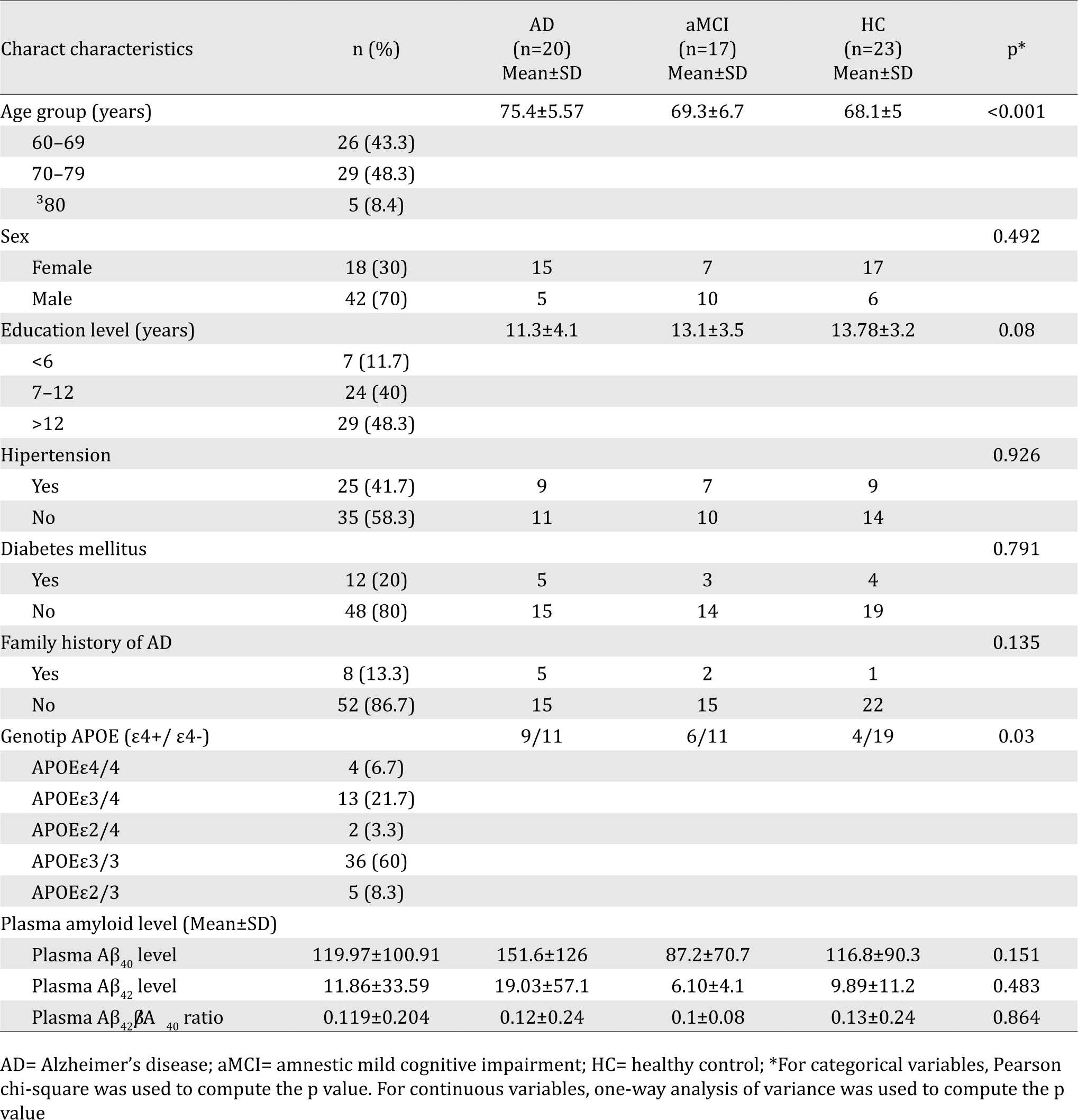
A total of 80 subjects in memory clinic were evaluated in which 65 patients among them met our inclusion criteria. We excluded five subjects due to a major infarct in MRI findings (one subject) and the impairment of renal function (four subjects). Sixty subjects were eligible for further step analyzing apo-E polymorphism and plasma amyloid beta (Aβ40 and Aβ42 level). Diagnosis is divided into three groups: AD = 20, aMCI = 17, and normal cognition = 23 subjects. Forty two subjects were women (70%) and unlike common demographic profile in Indonesia, half of the subjects had high educational level (>12 years of formal education) (Table 1).
Table 1. Subject characteristics
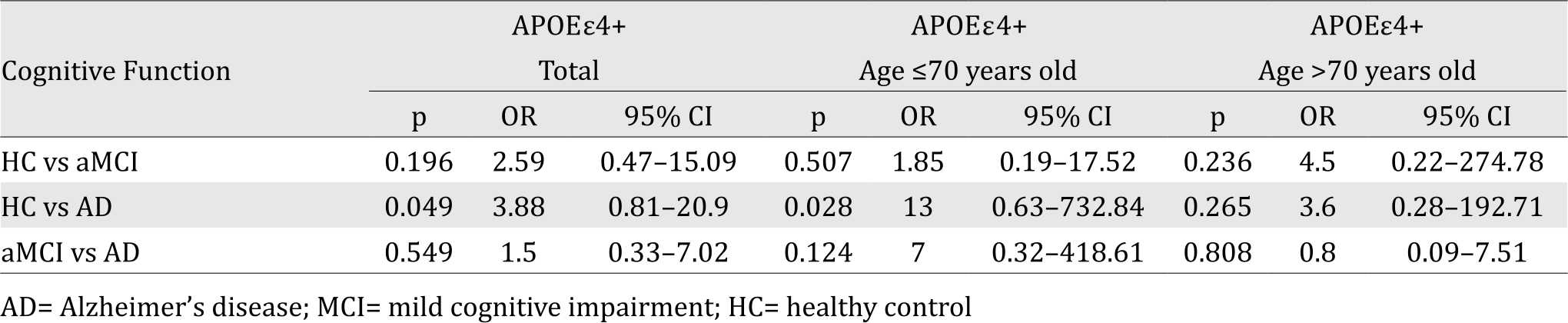
There is a significant association between APOEe4 genotype with cognitive function, particularly in HC versus AD group (Table 2). Subjects carrying the ε4 allele were more likely to have AD by 3.9 times compared to non-carriers. This likelihood is even higher in subjects with age-group ≤70 years (OR=13).
Table 2. Correlation between APOEε4 genotype with cognitive function
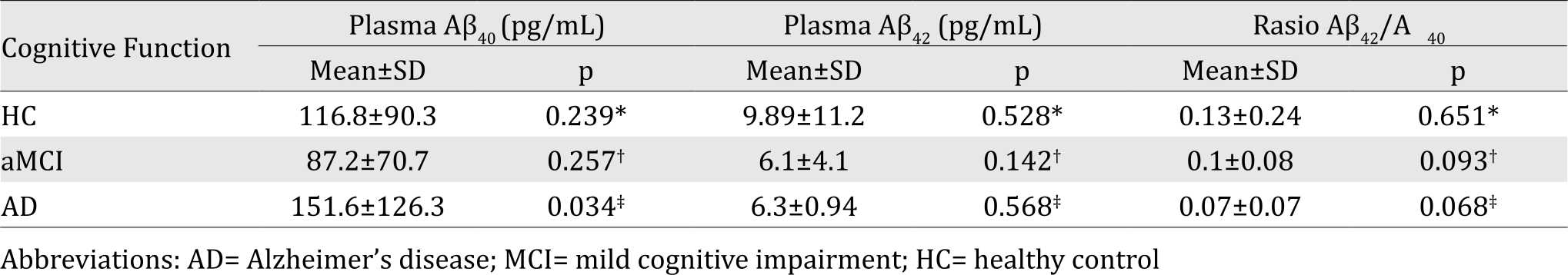
Level of Aβ40 and Aβ42 plasma in AD is the highest compared to aMCI and HC, while the Aβ42/Aβ40 ratio in this group is the lowest (Table 3). We found a significant difference between the mean of plasma Aβ40 in HC versus AD group (p<0.05).
Table 3. Mean difference of plasma Aβ in each cognitive function group
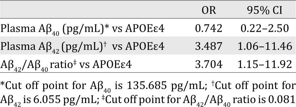
We analyzed the mean difference of plasma Aβ level in APOEε4(+) carrier compared to noncarrier and found it significant in level of Aβ42 plasma (Table 4). When we divided the subjects into two groups (higher level of Aβ and lower level of Aβ) based on certain cut off points, we found significant correlation between APOEε4(+) and higher Aβ42 plasma level (Table 5).
Table 4. Mean difference in subjects with APOEε4+ and APOEε4-
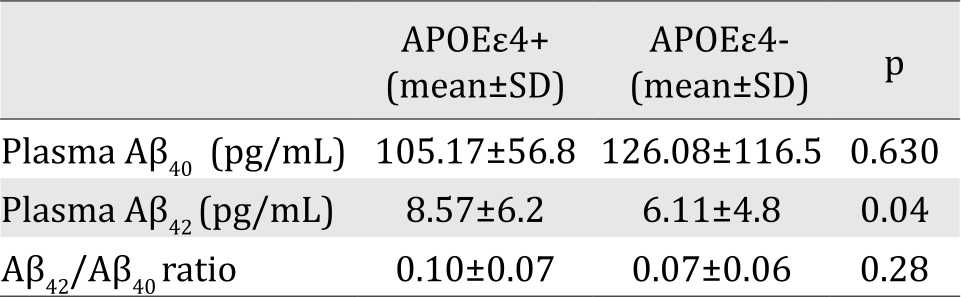
Table 5. Correlation between plasma Aβ and APOEε4
DISCUSSION
The ε4 allele of APOE as the strongest genetic risk factor for AD has been confirmed by genomewide association studies.33,34 The presence of this allele is associated with increased risk for both early-onset AD and late-onset AD.35,36 To our knowledge, investigation about the role of APOE ε4 in AD incidence among Indonesian has not been established. In population-based studies, the APOE ε4–AD association was weaker among African Americans (ε4/ε4, OR=5.7) and Hispanics (ε4/ε4, OR=2.2) and was stronger in Japanese people (ε4/ε4, OR=33.1) compared with Caucasian cases (ε4/ε4, OR=12.5).37 In this study, the role of APOEε4 as strong genetic risk factor had been proven; showing an increased risk of up to fourfold for carrier APOEε4 carriers to have AD than non-carriers. This risk is increased into 13-fold for subjects with age group ≤70 years, indicating that APOEε4 confers dramatically increased risk of development of AD with an earlier age of onset.
The investigation of plasma Aβ species offers advantage over conventional method for measuring Aβ level in the brain and CSF. Obtaining and analyzing plasma level is relatively inexpensive, minimally invasive, and can easily be performed at multiple time points. Therefore, a plasma-based biomarker for early detection and diagnosis of AD would be ideal. The utility of plasma Aβ as a potential AD biomarker had been assessed in previous studies, but the results have been inconsistent.38-41 It is well established that CSF Aβ42 level decreased in conjunction with the cognitive decline, it has been postulated that plasma Aβ42 may decrease similarly.42 Low plasma Aβ42 at baseline was associated with cognitive decline occurring during follow up.43 We found no statistical significance of differences in plasma Aβ42 levels and Aβ42/Aβ40 ratio among AD, MCI, and HC groups, but there is a tendency that both are lower in aMCI and AD subjects than in HC subjects. Interestingly, plasma Aβ40 is much higher in AD subjects compared to HC and aMCI subjects. Previous studies showed that Aβ levels varied highly in brains of patients with AD, and those with massive amyloid deposition contained predominantly Aβ40. In some patients with sporadic AD, high plasma levels of Aβ40 may contribute to the accumulation of Aβ40 in preexisting plaques.44
We found a significant correlation between APOEε4+ and high plasma Aβ42 level which more prominent in aMCI and AD groups. The APOEε4 has been associated with reduced Aβ clearance from the brain45 and plasma46 and with impaired tight junction integrity.47 Our data suggest that APOEε4 influences Aβ42 levels, but not Aβ40 level. Some studies have shown association between ε4 allele with increased fibrillar Aβ22 and decreased soluble plasma Aβ42 in a dosedependent manner.30 Discrepancies between our results and those reported by others may result from differences in patient populations, the use of different enzyme-linked immunosorbent assays and platforms, and timing of sample collection in relation to the stage of disease progression.
In conclusion, work summarized in this research highlights evidence for the association between APOEε4, plasma Aβ level, and cognitive decline. APOEε4 is a strong genetic risk factor for AD among Indonesian people, particularly in age group ≤70 years. APOEε4 seems to accelerate the rate and progression of AD through Aβ- dependent pathways. The exact mechanisms by which APOEε4 regulates Aβ aggregation and deposition require further investigation.
Conflicts of Interest
The authors affirm no conflict of interest in this study.
Acknowledgment
The authors would like to thank contributors who collected samples as well as patients and their families, whose participations and help make this work possible.
REFERENCES
- Alzheimer’s Association. 2012 Alzheimer’s disease facts and figures. Alzheimers Dement. 2012;8(2):131–68.
- Hardy J, Selkoe DJ. The amyloid hypothesis of Alzheimer’s disease: progress and problems on the road to therapeutics. Science. 2002;297(5580):353-6.
- Ballard C, Gauthier S, Corbett A, Brayne C, Aarsland D, Jones E. Alzheimer’s disease. Lancet. 2011;377(9770):1019–31.
- Corder EH, Saunders AM, Strittmatter WJ, Schmechel DE, Gaskell PC, Small GW, et al. Gene dose of apolipoprotein E type 4 allele and the risk of Alzheimer’s disease in late onset families. Science. 1993;261(5123):921–3.
- Bu G. Apolipoprotein E and its receptors in Alzheimer’s disease: pathways, pathogenesis and therapy. Nat Rev Neurosci. 2009;10(5):333–44.
- Huang Y, Mucke L. Alzheimer mechanisms and therapeutic strategies. Cell. 2012;148(6):1204–22.
- Farrer LA, Cupples LA, Haines JL, Hyman B, Kukull WA, Mayeux R, et al. Effects of age, sex, and ethnicity on the association between apolipoprotein E genotype and Alzheimer disease. A meta-analysis. APOE and Alzheimer Disease Meta Analysis Consortium. JAMA. 1997;278(16):1349–56.
- Rebeck GW, Reiter JS, Strickland DK, Hyman BT. Apolipoprotein E in sporadic Alzheimer’s disease: allelic variation and receptor interactions. Neuron. 1993;11(4):575–80.
- Morris JC, Storandt M, Miller JP, McKeel DW, Price JL, Rubin EH, et al. Mild cognitive impairment represents early-stage Alzheimer disease. Arch Neurol. 2001;58(3):397–405.
- Petersen RC, Smith GE, Waring SC, Ivnik RJ, Tangalos EG, Kokmen E. Mild cognitive impairment: clinical characterization and outcome. Arch Neurol. 1999;56(3):303–8.
- Smith GE, Bohac DL, Waring SC, Kokmen E, Tangalos EG, Ivnik RJ, et al. Apolipoprotein E genotype influences cognitive ‘phenotype’ in patients with Alzheimer’s disease but not in healthy control subjects. Neurology.1998;50(2):355–62.
- Farlow MR, He Y, Tekin S, Xu J, Lane R, Charles HC. Impact of APOE in mild cognitive impairment. Neurology. 2004;63(10):1898–901.
- Ramakers IH, Visser PJ, Aalten P, Bekers O, Sleegers K, van Broeckhoven CL, et al. The association between APOE genotype and memory dysfunction in subjects with mild cognitive impairment is related to age and Alzheimer pathology. Dement Geriatr Cogn Disord. 2008;26(2):101–8.
- Dik MG, Jonker C, Bouter LM, Geerlings MI, van Kamp GJ, Deeg DJ. APOE-epsilon4 is associated with memory decline in cognitively impaired elderly. Neurology. 2000;54(7):1492–7.
- Whitehair DC, Sherzai A, Emond J, Raman R, Aisen PS, Petersen RC, et al. Influence of apolipoprotein E varepsilon4 on rates of cognitive and functional decline in mild cognitive impairment. Alzheimers Dement. 2010;6(5):412–9.
- Cosentino S, Scarmeas N, Helzner E, Glymour MM, Brandt J, Albert M, et al. APOE epsilon 4 allele predicts faster cognitive decline in mild Alzheimer disease. Neurology. 2008;70(19Pt2):1842–9.
- Fleisher AS, Sowell BB, Taylor C, Gamst AC, Petersen RC, Thal LJ, et al. Clinical predictors of progression to Alzheimer disease in amnestic mild cognitive impairment. Neurology. 2007;68(19):1588–95.
- Elias-Sonnenschein LS, Viechtbauer W, Ramakers IH, Verhey FR, Visser PJ. Predictive value of APOE-ε4 allele for progression from MCI to AD-type dementia: a meta-analysis. J Neurol Neurosurg Psychiatry. 2011;82(10):1149–56.
- Petersen RC, Smith GE, Ivnik RJ, Tangalos EG, Schaid DJ, Thibodeau SN, et al. Apolipoprotein E status as a predictor of the development of Alzheimer’s disease in memoryimpaired individuals. JAMA. 1995;273(16):1274–8.
- Vemuri P, Wiste HJ, Weigand SD, Knopman DS, Shaw LM, Trojanowski JQ, et al. Effect of apolipoprotein E on biomarkers of amyloid load and neuronal pathology in Alzheimer disease. Ann Neurol. 2010;67(3):308–16.
- Liu CC, Kanekiyo T, Xu H, Bu G. Apolipoprotein E and Alzheimer disease: risk, mechanisms and therapy. Nat Rev Neurol. 2013;9(2):106–18.
- Reiman EM, Chen K, Liu X, Bandy D, Yu M, Lee W, et al. Fibrillar amyloid-beta burden in cognitively normal people at 3 levels of genetic risk for Alzheimer’s disease. Proc Natl Acad Sci U S A. 2009;106(16):6820–5.
- Namba Y, Tomonaga M, Kawasaki H, Otomo E, Ikeda K. Apolipoprotein E immunoreactivity in cerebral amyloid deposits and neurofibrillary tangles in Alzheimer’s disease and kuru plaque amyloid in Creutzfeldt-Jakob disease. Brain Res. 1991;541(1):163–6.
- Kok E, Haikonen S, Luoto T, Huhtala H, Goebeler S, Haapasalo H, et al. Apolipoprotein E-dependent accumulation of Alzheimer disease-related lesions begins in middle age. Ann Neurol. 2009;65(6):650–7.
- Polvikoski T, Sulkava R, Haltia M, Kainulainen K, Vuorio A, Verkkoniemi A, et al. Apolipoprotein E, dementia, and cortical deposition of beta-amyloid protein. N Engl J Med. 1995;333(19):1242–7.
- Schmechel DE, Saunders AM, Strittmatter WJ, Crain BJ, Hulette CM, Joo SH, et al. Increased amyloid betapeptide deposition in cerebral cortex as a consequence of apolipoprotein E genotype in late-onset Alzheimer disease. Proc Natl Acad Sci U S A. 1993;90(20):9649–53.
- Head D, Bugg JM, Goate AM, Fagan AM, Mintun MA, Benzinger T, et al. Exercise engagement as a moderator of the effects of APOE genotype on amyloid deposition. Arch Neurol. 2012;69(5):636–43.
- Prince JA, Zetterberg H, Andreasen N, Marcusson J, Blennow K. APOE epsilon4 allele is associated with reduced cerebrospinal fluid levels of Abeta42. Neurology. 2004;62(11):2116–8.
- Swaminathan S, Risacher SL, Yoder KK, West JD, Shen L, Kim S, et al. Association of plasma and cortical amyloid beta is modulated by APOEε4 status. Alzheimers Dement. 2014;10(1):e9–18.
- Toledo JB, Vanderstichele H, Figurski M, Aisen PS, Petersen RC, Weiner MW, et al. Factors affecting Ab plasma levels and their utility as biomarkers in ADNI. Acta Neuropathol. 2011;122(4):401–13.
- Lui JK, Laws SM, Li QX, Villemagne VL, Ames D, Brown B, et al. Plasma amyloid-beta as a biomarker in Alzheimer’s disease: the AIBL study of aging. J Alzheimers Dis. 2010;20(4):1233–42.
- Devanand DP, Schupf N, Stern Y, Parsey R, Pelton GH, Mehta P, et al. Plasma Ab and PET PiB binding are inversely related in mild cognitive impairment. Neurology. 2011;77(2):125–31.
- Bunce D, Fratiglioni L, Small BJ, Winblad B, Bäckman L. APOE and cognitive decline in preclinical Alzheimer disease and non-demented aging. Neurology. 2004;63(5):816-21.
- Lin AL, Laird AR, Fox PT, Gao JH. Multimodal MRI neuroimaging biomarkers for cognitive normal adults, amnestic mild cognitive impairment, and Alzheimer’s disease. Neurol Res Int. 2012;2012:907409.
- Caselli RJ, Reiman EM, Locke DE, Hutton ML, Hentz JG, Hoffman-Snyder C, et al. Cognitive domain decline in healthy apolipoprotein E epsilon4 homozygotes before the diagnosis of mild cognitive impairment. Arch Neurol. 2007;64(9):1306–11.
- Caselli RJ, Reiman EM, Osborne D, Hentz JG, Baxter LC, Hernandez JL, et al. Longitudinal changes in cognition and behavior in asymptomatic carriers of the APOE e4 allele. Neurology. 2004;62(11):1990–5.
- Caselli RJ, Dueck AC, Osborne D, Sabbagh MN, Connor DJ, Ahern GL, et al. Longitudinal modeling of age-related memory decline and the APOE epsilon4 effect. N Engl J Med. 2009;361(3):255–63.
- Mayeux R, Schupf N. Blood-based biomarkers for Alzheimer’s disease: plasma Ab40 and Ab42, and genetic variants. Neurobiol Aging. 2011;32(Suppl1):S10–9.
- Song F, Poljak A, Valenzuela M, Mayeux R, Smythe GA, Sachdev PS. Meta-analysis of plasma amyloid-b levels in Alzheimer’s disease. J Alzheimers Dis. 2011;26(2):365–75.
- Koyama A, Okereke OI, Yang T, Blacker D, Selkoe DJ, Grodstein F. Plasma amyloid-b as a predictor of dementia and cognitive decline: a systematic review and meta-analysis. Arch Neurol. 2012;69(7):824–31.
- Thambisetty M, Lovestone S. Blood-based biomarkers of Alzheimer’s disease: challenging but feasible. Biomark Med. 2010;4(1):65–79.
- Graff-Radford NR, Crook JE, Lucas J, Boeve BF, Knopman DS, Ivnik RJ, et al. Association of low plasma Abeta42/ Abeta40 ratios with increased imminent risk for mild cognitive impairment and Alzheimer disease. Arch Neurol. 2007;64(3):354–62.
- Seppälä TT, Herukka SK, Hänninen T, Tervo S, Hallikainen M, Soininen H, et al. Plasma Abeta42 and Abeta40 as markers of cognitive change in follow-up: a prospective, longitudinal, population-based cohort study. J Neurol Neurosurg Psychiatry. 2010;81(10):1123–7.
- Ishii K, Tamaoka A, Mizusawa H, Shoji S, Ohtake T, Fraser PE, et al. Abeta1–40 but not Abeta1–42 levels in cortex correlate with apolipoprotein E epsilon4 allele dosage in sporadic Alzheimer’s disease. Brain Res. 1997;748(1– 2):250–2.
- Castellano JM, Kim J, Stewart FR, Jiang H, DeMattos RB, Patterson BW, et al. Human apoE isoforms differentially regulate brain amyloid-b peptide clearance. Sci TransI Med. 2011;3(89):89ra57.
- Sharman MJ, Morici M, Hone E, Berger T, Taddei K, Martins IJ, et al. APOE genotype results in differential effects on the peripheral clearance of amyloid-beta42 in APOE knock-in and knock-out mice. J Alzheimers Dis. 2010;21(2):403–9.
- Nishitsuji K, Hosono T, Nakamura T, Bu G, Michikawa M. Apolipoprotein E regulates the integrity of tight junctions in an isoform-dependent manner in an in vitro blood-brain barrier model. J Biol Chem. 2011;286(20):17536–42.
Copyright @ 2016 Authors. This is an open access article distributed under the terms of the Creative Commons Attribution-NonCommercial 4.0 International License (http://creativecommons.org/licenses/by-nc/4.0/), which permits unrestricted non-commercial use, distribution, and reproduction in any medium, provided the original author and source are properly cited.
mji.ui.ac.id