Section Abstract Introduction Methods Results Discussion Conflict of Interest Acknowledgment References
Basic Medical Research
The suppression of manganese superoxide dismutase decreased the survival of human glioblastoma multiforme T98G cells
pISSN: 0853-1773 • eISSN: 2252-8083
http://dx.doi.org/10.13181/mji.v26i1.1511 Med J Indones. 2017;26:19–25
Received: July 26, 2016
Accepted: January 29, 2017
Author affiliation:
1 Department of Biochemistry and Molecular Biology, Faculty of Medicine, Universitas Indonesia, Jakarta, Indonesia
2 Center of Hypoxia and Oxidative Stress Studies (CHOSS), Faculty of Medicine, Universitas Indonesia, Jakarta, Indonesia
3 Department of Anatomic Pathology, Faculty of Medicine, Universitas Indonesia, Jakarta, Indonesia
Corresponding author:
Septelia I. Wanandi
E-mail: septelia@gmail.com
Background
Glioblastoma multiforme (GBM) is a primary malignant brain tumor which has poor prognosis. High incidence of oxidative stress-based therapy resistance could be related to the high antioxidant status of GBM cells. Our previous study has reported that manganese superoxide dismutase (MnSOD) antioxidant expression was significantly higher in high grade glioma than in low grade. The aim of this study was to analyze the impact of MnSOD suppression toward GBM cell survival.
Methods
This study is an experimental study using human glioblastoma multiforme T98G cell line. Suppression of MnSOD expression was performed using in vitro transfection MnSOD-siRNA. The MnSOD expression was analyzed by measuring the mRNA using real time RT-PCR, protein using ELISA technique, and specific activity of enzyme using inhibition of xantine oxidase. Concentration of reactive oxygen species (ROS) intracellular was determined by measuring superoxide radical and hydrogen peroxide. Cell survival was analyzed by measuring viability, proliferation, and cell apoptosis.
Results
In vitro transfection of MnSOD-siRNA suppressed the mRNA, protein, and specific activity of MnSOD. This treatment significantly increased the concentration of superoxide radical; however, it did not influence the concentration of hydrogen peroxide. Moreover, viability MnSOD-suppressing cell significantly decreased, accompanied by increase of cell apoptosis without affecting cell proliferation.
Conclusion
The suppression of MnSOD expression leads to decrease glioblastoma multiforme cell survival, which was associated to the increase of cell apoptotic.
Keywords
Cell survival, glioblastoma multiforme, MnSOD-siRNA, ROS, T98G cells
Glioblastoma multiforme is the most malignant primary brain tumor which arises from glial cells. Glioblastoma multiforme patients have poor prognosis due to the resistance and the recurrence of the conventional treatment which encompasses open craniotomy surgery followed by radiotherapy or chemotherapy.1 Radiotherapy kills cancer cells through reactive oxygen species which causes oxidative damage.2 However, it can be prevented through the action of endogenous antioxidant enzyme by eliminating reactive oxygen species (ROS).3 Therefore, endogenous antioxidant enzyme has an essential contribution for the success of the treatment.
The main endogenous antioxidant enzyme is manganese superoxide dismutase (MnSOD) which is located in mitochondrial matrix. This enzyme eliminates superoxide radical (O2) by catalyzing the changeover of O2 to oxygen (O2) and hydrogen peroxide (H2O2).3 Several studies showed that the up-regulation of MnSOD in some cancers including glioma was correlated with poor tumor prognosis.4–6 Moreover, MnSOD has been observed as an anti-apoptotic molecule, and increasing of MnSOD in tumor could lead to treatment resistance and tumor metastatasis.7–9 Our previous study using glioma tissues from glioma patients showed that the expression of MnSOD mRNA was higher in high grade glioma than in low grade glioma and it was associated with the increase of oxidative defects. Moreover, our research proved that the expression of MnSOD mRNA was correlated with the tumor grade.10 Regarding that problem, we intend to further explore the role of MnSOD expression in high grade glioma cell survival through the suppression of MnSOD in human glioblastoma multiforme T98G cell line as the highest grade glioma. The aim of this study is to analyze the impact of MnSOD suppression on intracellular ROS, cell proliferation, cell viability, and apoptosis.
METHODS
The human glioblastoma multiforme T98G cell line (American type culture collection (ATCC) Number CRL-1690TM) used in this experimental study was kindly provided by Prof. Alexander Brehm from the Institut fuer Molekularbiologie und Tumorforschung Philipps Universitaet Marburg, Germany. T98G cell line was used because this cell line is a suitable transfection host according to ATCC general information. Our study was conducted in the Laboratory of Institute Human Virology and Cancer Biology Universitas Indonesia (IHVCB-UI), the Makmal Terpadu Faculty of Medicine Universitas Indonesia (FMUI), Biochemistry & Molecular Biology FMUI, and the Eijkman Institute.
Cell culture
The T98G human glioblastoma cell line was grown on T-25 cm2 plastic tissue culture flasks (Corning) in high glucose Dulbecco’s Modified Eagle’s Medium supplemented with 3.7 g/L of sodium bicarbonate, 10% heat-inactivated fetal bovine serum (Biowest), 1% streptomycin - penicillin, and 1% amphotericine at 37°C in a humidified atmosphere of 95% air and 5% CO2. Subcultivation of the confluence cells were done using 0.25% trypsin and 1% ethylenediaminetetraacetic acid (EDTA). The medium of cell cultures was replaced every three days. MnSOD small interfering ribonucleic acid (siRNA) transfection utilized fifty thousand T98G cells which were grown in triplicated 24-well plates. The treatment was repeated three times at different times.
MnSOD-siRNA transfection
MnSOD siRNA sequences (Custom Synthesized siRNA, Ambion) were 5’-GGAACAACAGGCCUUAUUCTT-3’ (sense) and 5’-GAAUAAGGCCUGUUGUUCCTT-3’ (antisense).11 Silencer select negative control number 1 siRNA (Ambion) was used as a negative control. Fifty thousand T98G cells were grown in triplicated 24-well plates and transfected with either 60 nM of MnSOD-siRNA or 30 nM of the negative control siRNA in serum-free medium (OptiMEM I, Gibco) by using Lipofectamine reagent 2,000 (Invitrogen®) according the manufacture’s instruction. In the following two days, the cells were washed, trypsinized, and extracted for measurement of MnSOD expression, intracellular ROS, cell viability, cell proliferation, and apoptosis.
Analysis of MnSOD mRNA using real time RTPCR
Ribonucleic acid (RNA) was isolated from the T98G cells using Tripure RNA Isolation Kit (Roche®). RNA templates were amplified using iScript one step real-time polymerase chain reaction (RT-PCR) Kit with SYBR Green (BioRad). Reaction protocols was described as our previous research.12 MnSOD gene primer were GCACTAGCAGCATGTTGAGC (forward) and ACTTCTCCTCGGTGACGTTC (reverse) with product size 216 bp. Housekeeping gene primer (18S rRNA) were AAACGGCTACCACATCCAAG (forward) and CCTCCAATGGATCCTCGTTA (reverse) with product size 155 bp. Relative expression of MnSOD mRNA in T98G cells posttransfection was calculated using Livax formula and normalized against T98G cells without the treatment as a control.
Analysis of MnSOD protein
Analysis of MnSOD protein was performed using enzyme linked immunosorbent assay (ELISA) technique (NWLSSTM MnSOD ELISA kit) in accordance with manufacturer’s protocol. Protein isolated with Tripure Isolation Reagent was used for this assay. The human MnSOD standard (10 ng/mL) was reconstituted by adding 1 mL of standard buffer into the standard protein bottle comprising lyophilized human MnSOD protein. One hundred μL of diluted standards, 100 μL of sample dilution buffer, and 100 μL of sample were added to the appropriate microtiter wells according to plan. The MnSOD samples were determined by comparing their absorbance at 450 nm with the standard curve.
Analysis of MnSOD specific enzyme activity
Analysis of MnSOD specific enzyme activity was detected using RanSOD® kit according to manufacturer’s protocol. Inhibition of Cu/ZnSOD was performed by adding sodium cyanide (5 mM) into each sample, followed by incubation for five minutes at room temperature. The percentage of inhibition from each samples were plotted to the standard curve to obtain the enzyme activity calculation. The enzyme activity (in units) per mg protein represented the specific activity of MnSOD enzyme. Concentration of protein was detected based on the plotting of samples absorbance at 280 nm to the bovine serum albumin (BSA) standard curve.12
Determination of ROS
Analysis of intracellular hydrogen peroxide using DCFH-DA
Hydrogen peroxide was analyzed by measuring the 2’,7’-dichlorodihydrofluorescein diacetate (DCFH-DA, molecular probes, USA). Intracellular peroxide would oxidize DCFH-DA to form the fluorescent compound 2’,7’-dichlorofluorescein (DCF). Fifty thousand T98G cells were washed and centrifuged to obtain cell pellets. Cell pellets were added to 20 μM of DCFH-DA in 500 μL PBS and incubated for 30 minutes at 37°C in the dark. The cell pellets were rinsed with PBS (2X), and intensity of fluorescence was detected using a spectrofluorometer (Perkin Elmer) with excitation at 485 nm and emission at 530 nm.13
Analysis of intracellular superoxide anion by dihydroethidium (DHE)
Dihydroethidium (DHE) was oxidized to ethidium in the presence of superoxide anion. Fresh Dihydroethidium/DHE (invitrogen) solutions were prepared by dissolving 2 μL of DHE stock solution (5mM) in 500 μL PBS, so the final concentration of DHE was 20 μM. Solutions containing DHE were protected from light before and during the experiments. Twenty thousand T98G cells were washed and centrifuged to obtain cell pellets. Cell pellets were diluted in PBS containing 20 μM of DHE and incubated for 30 minutes at 37°C. The cells were washed with PBS (2x) and intensity of fluorescence was detected by a spectrofluorometer with excitation at 488 nm and emission at 585 nm.14
Cell viability using MTS assay (The CellTiter 96® Aqueous Assay, Promega)
The CellTiter 96® Aqueous Assay is consists of solutions of a novel tetrazolium substance [3-(4,5-dimethylthiazol-2-yl)-5-(3-carboxymethoxyphenyl)-2-(4-sulfophenyl)-2Htetrazolium, inner salt; MTS(a)] and an electron coupling reagent (phenazine methosulfate; PMS). Ten thousand cells after MnSOD siRNA transfection were washed, and 120 μL of the combined MTS/PMS solution in PBS into each well were added. The plate was incubated for one hour at 37°C in a humidified, 5% CO2. Cell viability was detected by measuring the formazan product at 490 nm absorbance which was proportional to the total sum of viable cells in culture.
Analysis of cell proliferation
T98G cell proliferation was analyzed using The Cell Proliferation ELISA, BrdU kit (Roche®) according to manufacturer’s protocol. Twenty thousand cells after MnSOD siRNA transfection were added with 20 μL/well of BrdU and incubated for 24 hours. In this process, the BrdU as the pyrimidine analogue was integrated into the DNA of proliferating cells. The developed color was measured using ELISA reader at 490 nm absorbance. This absorbance numbers were directly associated with the sum of DNA synthesis and represent the number of cells proliferation.
Analysis of cytochrome c
After transfection, cells (1x10⁵) were washed, trypsinized, and centrifuged to obtain cell pellets. The cell pellets were diluted in 300 μL of cell lysis buffer and incubated for one hour at room temperature, then they were centrifuged at 1,000x g for 15 minutes and aspirated the supernatant for cytochrome c measurement (Human Cytochrome c Quantikine ELISA kit, R&D System®) in accordance with the manufacturer’s protocol.
TUNEL analysis
The apoptosis was analyzed by measuring deoxyribonucleic acid (DNA) fragmentation using terminal deoxynucleotydil transferase dUTP nick end labeling (TUNEL) technology (in situ Cell Death Detection kit, POD) according to the manufacturer’s protocol. Fifty thousand T98G cells were plated in slide chamber one day before transfection. Post-transfection, the T98G cells were washed and fixed in 100 μL of fresh 4% para formaldehyde in PBS pH 7.4, then they were incubated for one hour at a room temperature. In addition, methyl green solution (2.5 mg in 500 μL buffer) was used in the counterstaining process. The apoptotic cells were characterized by the brown cells, which were then calculated by using statistical software cell counter.
Statistical analysis
All the treatment samples were compared and divided to control cells to get the ratio value, which were presented as a mean. The difference of statistical significance between groups was analyzed by the Student’s t test and Wilcoxon test using predictive analytics software (PASW) statistic 18 software. The statistical significance was set at p<0.05.
RESULTS
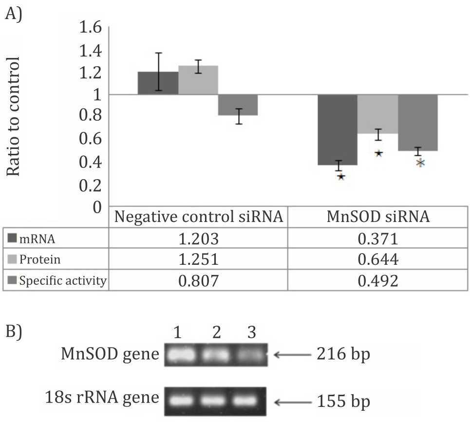
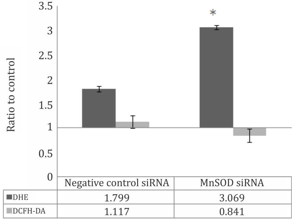
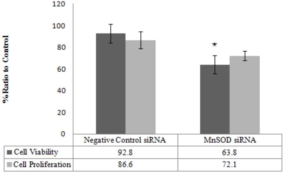
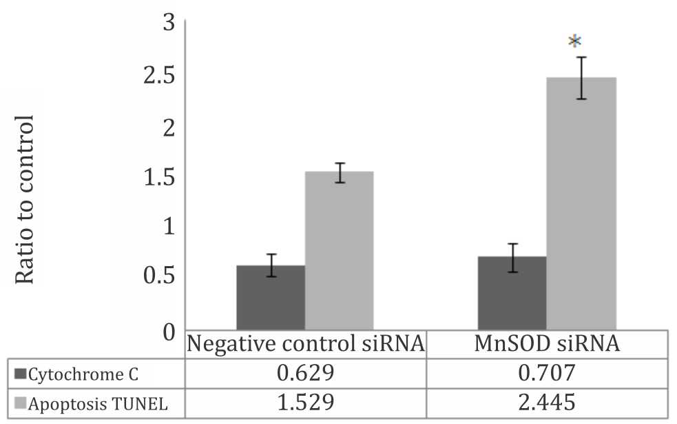
The in vitro transfection of MnSOD siRNA inhibited the MnSOD expression not only at the level of mRNA, but also at the protein and specific activity (Figure 1A). The electrophoresis of real time RT PCR product showed that the band intensity of MnSOD-siRNA cells was shown to decrease compared to the control (Figure 1B). Obviously, this treatment significantly increased superoxide radical and slightly decreased hydrogen peroxide (Figure 2). Suppression of MnSOD expression in our research significantly reduced the cells viability (Figure 3), even significantly increased cells apoptosis (Figure 4). Cell proliferation also slightly decreased in MnSOD-siRNA transfected T98G cells; however, it was not statistically significant (Figure 3). Cytochrome c slightly increased in MnSOD-siRNA transfected T98G cells, but was not statistically significant (Figure 4). Figure 5 shows the microphotograph results of cells apoptosis using TUNEL technique.

Figure 1. MnSOD expression was presented as the ratio to control. The control was T98G cells without any treatment. A) In vitro MnSOD-siRNA transfection significantly decreased not only at MnSOD mRNA level around 62.9% reduction (*p<0.05), but also at protein around 35.6% reduction (*p<0.05) and specific activity around 50.8% reduction (*p<0.01); B) Electrophoresis reaL time RT PCR product: 1). Cells; 2). Cells + siRNA negative control; 3). Cells + MnSOD siRNA

Figure 2. Intraselullar reactive oxygen species (ROS) was presented as the ratio to control. The control was T98G cells without any treatment. Dihydroethidium (DHE) reflected superoxide radical significantly (*p<0.01) increased in MnSODsuppressing cells compared to the negative control

Figure 3. Cell viability and cell proliferation were presented as the percentage of ratio to control. The control was T98G cells without any treatment. Cell viability was significantly lower in MnSOD-suppressing cells (*p<0.05) compared to the negative control. MnSOD-suppressing cell proliferation was slightly decreased, but it is not statistically significant

Figure 4. Apoptosis and cytochrome c were presented as the ratio to control. The control was T98G cells without any treatment. Fragmentation DNA by TUNEL analysis significantly increased in MnSOD-suppressing cell (∗p<0.01) compared to the negative control. Cytochrome C slightly increased in MnSOD-suppressing cells but it is not significant

Figure 5. Microscopic TUNEL results (200X magnification). Apoptotic cells were characterized by d ark brown cells A) positive control (cells + DNAse); B) negative control (cells + PBS); C) cells control (without treatment); D) cells + negative control siRNA; E) Cells + MnSOD siRNA
DISCUSSION
Our previous study showed that MnSOD mRNA expression in the high grade glioma cells from the glioma patients significantly increased compared to the low grade one.10 Several other studies showed that high expression of MnSOD in some cancer cells were correlated with resistance to the treatment, metastasis, and poor prognosis.4–9,15 Reddy et al16 identified several genes that regulate the degree of malignancy in glioblastoma multiforme. One of them is MnSOD that increased in most of primary GBM. Therefore, to further analyze the role of MnSOD in the high grade glioma, we suppressed the MnSOD mRNA expression using in vitro MnSOD-siRNA transfection. Indeed, glioblastoma multiforme T98G cell line was used for this research since the amount limitation of the tissues samples from glioma patient made it difficult to perform primary culture. We demonstrated that the in vitro transfection of MnSOD siRNA inhibited the MnSOD expression not only at the level of mRNA, but also at level of protein and specific activity.
MnSOD catalyzes the dismutation of superoxide anion into hydrogen peroxide and water molecule.3 Therefore, superoxide radical will increase when MnSOD is suppressed, as shown in our result. The inhibition of MnSOD mRNA expression obviously increases the DHE that reflects superoxide radical.17 On other hand, hydrogen peroxide as a product of MnSOD conversion will be decreased because the accumulation of superoxide radical is not converted into hydrogen peroxide due to lack of MnSOD. That reason was proved in our results that DCFH-DA as a selective marker for hydrogen peroxide, lipid peroxidation, and hydroxyl17 decreased in MnSOD-siRNA transfected cells. Teoh et al18 also demontrated that DCFH-DA in MnSOD-suppressing pancreatic cells was lower than in the negative control, which was similar with our results.
The suppression of MnSOD expression significantly decreased the survival rates of T98G cells which was identified by the decreasing cells viability and the increasing apoptosis. Similar result was also found by Kuninaka et al19 which showed that cell viability in the MnSODsuppressing colorectal cancer cells was lower than in the negative control. Moreover, the study demonstrated that suppression of MnSOD activity on colon cancer cells increased the apoptosis induced by radiation, hyperthermia, and doxorubicin drug. Otherwise, upregulation of MnSOD in esophageal cancer cells was associated with apoptosis resistance.9 Mechanism of increasing apoptosis in MnSOD-suppressing cells was probably due to the increasing of superoxide radical. Hileman et al20 suggested that SOD inhibition could be used for inhibiting the grow rate of cancer cells due to accumulation of superoxide radical that cause cell death.
The augmentation of ROS leads to increase the permeability of mitochondria outer membrane. Therefore, Cytochrome c will be released from mitochondria inter-membrane space into cytosol. Cytochrome c binds with Apaf-1 forming apoptosome. Apoptosome formation will activate caspase-9, which causes cascade caspase activation to a cell death.21 Cell proliferation slightly decreased in MnSOD-suppressing cells, but it was not statistically significant. Decreased in MnSOD-suppressing T98G cell proliferation in our research probably was caused by superoxide radical accumulation. That condition may activate the JNK signaling cascade thereby cell division stop.22 The increase of superoxide radical also inhibites human aortic endothelial and rat stellate cells.23–24 Nevertheless, the accumulation of superoxide radical in this research clearly causes more apoptosis than in cells proliferation inhibition.
This result is still limited at the level in vitro study which has not yet represented the real of high grade glioma condition. Nevertheless, the impact of this research has promising therapeutic potential for glioma patients. Indeed, further studies are required with in vivo MnSOD-siRNA transfection in animals with glioblastoma multiforme to ensure the role of MnSOD on glioma cell survival as the early step to do MnSOD-siRNA transfection for the therapy of glioma patients. Probably, intra-tumor transfection by injecting to the tumor site directly can be chosen to make it selective for tumor cells.
We conclude that the suppression of MnSOD expression causes a significantly decreased glioblastoma multiforme T98G cell survival associated with the increase of cell apoptotic.
Conflicts of Interest
The authors affirm no conflict of interest in this study.
Acknowledgment
This research was funded by Riset Unggulan Universitas Indonesia (RUUI) 2010. The authors would like to express their gratitude to Direktorat Riset dan Pengabdian Masyarakat Universitas Indonesia (DRPM-UI) for supporting the research.
REFERENCES
- Walid MS. Prognostic factor for long-term survival after glioblastoma. Perm J. 2008;12(4):45–8.
- Azzam EI, Jay-Gerin JP, Pain D. Ionizing radiationinduced metabolic oxidative stress and prolonged cell injury. Cancer Lett. 2012;327(1–2):48–60.
- Halliwell B, Gutteridge JMC. Free radical in biology and medicine. 4th ed. London: Oxford University Press; 2007. p. 30–74.
- Dhar SK, Tangpong J, Chaiswing L, Oberley TD, St. Clair DK. Manganese superoxide dismutase is a p53- regulated gene that switches cancers between early and advanced staged. Cancer Res. 2011;71(21):6684–95.
- Kattan J, Minig V, Leroy P, Dauça M, Becuwe P. Role of manganese superoxide dismutase on growth and invasive properties of human estrogen-independent breast cancer cells. Breast Cancer Res Treat. 2008;108(2):203–15.
- Järvelä S, Bragge H, Paunu N, Järvelä T, Paljärvi H, Kalimo H, et al. Antioxidant enzymes in oligodendroglial brain tumors: association with proliferation, apoptotic activity and survival. J Neurooncol. 2006;77(2):131–40.
- Kamarajugadda S, Cai Q, Chen H, Nayak S, Zhu J, He M, et al. Manganese superoxide dismutase promotes anoikis resistance and tumor metastasis. Cell Death Dis. 2013;4:e504.
- Yeung BHY, Wong KY, Lin MC, Wong CKC, Mashima T, Tsuruo T, et al. Chemosensitisation by manganese superoxide dismutase inhibition is caspase-9 dependent and involves extracellular signal-regulated kinase I/2. Br J Cancer. 2008;99(2):283–93.
- Hu H, Luo LM, Du XL, Feng YB, Zhang Y, Shen XM, et al. Up-regulated manganese superoxide dismutase expression increases apoptosis resistance in human esophageal squamous cell carcinomas. Chin Med J. 2007;120(23):2092–8.
- Hardiany NS, Mulyawan W, Wanandi SI. Correlation between oxidative stress and tumor grade in glioma cells from patients in Jakarta. Med J Indones. 2012;21(3):122–7.
- Comhair SA, Xu W, Ghosh S, Thunnissen F, Almasan A, Calhoun WJ, et al. Superoxide dismutase inactivation in pathophysiology of asthamatic airway remodeling and reactivity. Am J Pathol. 2005;166(3):663–74.
- Hardiany NS, Sadikin M, Wanandi SI. Gene expression of manganese superoxide dismutase (MnSOD) in human glioma cells. Med J Indones. 2010;19(1):21–5.
- Mao W, Chen X, Yang T, Yin Y, Ge M, Luo M, et al. A rapid fluorescent screening method for cellular sensitivity to anti-cancer compound. Cytotechnology. 2012;64(4):451–7.
- Viola G, Salvador A, Vedaldi D, Fortunato E, Disaro S, Basso G, et al. Induction of apoptosis by photoexcited tetracyclic compounds derivatives of benzo[b] thiophenes and pyridines. J Photochemist Photobiol B: Biol. 2006;82(2):105–16.
- Hempel N, Carrico PM, Melendez JA. Manganese superoxide dismutase (Sod2) and redox-control of signaling events that drive metastasis. Anticancer Agents Med Chem. 2011;11(2):191–201.
- Reddy SP, Britto R, Vinnakota K, Aparna H, Sreepathi HK, Thota B, et al. Novel glioblastoma multiforme markers with diagnostic and prognostic value identified through transcriptome analysis. Clin Cancer Res. 2008;14(10):2978–87.
- Kalyanaraman B, Darley-Usmar V, Davies KJ, Dennery PA, Forman HJ, Grisham MB. Measuring reactive oxygen and nitrogen species with fluorescent probes: challenges and limitations. Free Radic Biol Med. 2012;52(1):1–6.
- Teoh ML, Sun W, Smith BJ, Oberley LW, Cullen JJ. Modulation of reactive oxygen species in pancreatic cancer. Clin Cancer Res. 2007;13(24):7441–50.
- Kuninaka S, Ichinose Y, Koja K, Toh Y. Suppression of manganese superoxide dismutase augments sensitivity to radiation, hyperthermia and doxorubicin in colon cancer cell lines by inducing apoptosis. Br J Cancer. 2000;83(7):928–34.
- Hileman EA, Achanta G, Huang P. Superoxide dismutase: an emerging target for cancer therapeutics. Expert Opin Ther Targets. 2001;5(6):697–710.
- Murphy BM, Seamus JM. Caspase structure, activation pathways and substrate. In: Yin XM, Dong Z, editors. Essentials of apoptosis. New Jersey: Humana Press, 2003; p. 3–9.
- Gerhard Kauss. Biochemistry of signal transduction and regulation. 2nd ed. Wiley-VCH 2001; p.351–6.
- Dunning S, Hannivoort RA, de Boer JF, Buist-Homan M, Faber KN, Moshage H. Superoxide anions and hydrogen peroxide inhibit proliferation of activated rat stellate cells and induce different modes of cell death. Liver Int. 2009;29(6):922–32.
- Zanetti M, Zwacka R, Engelhardt J, Katusic Z, O‘Brien T. Superoxide anion and endothelial cell proliferation in normoglycemia and hyperglycemia. Arterioscler Thromb Vasc Biol. 2001;21(2):195–200.
Copyright @ 2017 Authors. This is an open access article distributed under the terms of the Creative Commons Attribution-NonCommercial 4.0 International License (http://creativecommons.org/licenses/by-nc/4.0/), which permits unrestricted non-commercial use, distribution, and reproduction in any medium, provided the original author and source are properly cited.
mji.ui.ac.id