Section Abstract Introduction Methods Results Discussion Conflict of Interest Acknowledgment References
Basic Medical Research
Expressions of stemness markers in keloid tissue
pISSN: 0853-1773 • eISSN: 2252-8083
https://doi.org/10.13181/mji.v27i3.1920 Med J Indones. 2018;27:145–9
Received: March 21, 2017
Accepted: June 5, 2018
Author affiliation:
1 Master of Biomedical Sciences, Faculty of Medicine, Universitas Indonesia, Jakarta, Indonesia
2 Department of Histology, Faculty of Medicine, Universitas Indonesia, Jakarta, Indonesia
3 Department of Biochemistry and Molecular Biology, Faculty of Medicine, Center of Hypoxia and Oxidative Stress Studies, Universitas Indonesia, Jakarta, Indonesia
Corresponding author:
Sri W.A. Jusman
E-mail: sriwidiaaj@gmail.com
Background
Keloid is an abnormal wound healing process that extends beyond the site of injury. Keloid and tumor’s shared similarity of recurrence suggesting a shared underlying mechanism that involves stemness. Octamer-binding transcription factor-4 (Oct-4) and aldehyde dehydrogenase-1 (ALDH1) are stem cell stemness markers. This study aimed to analyze Oct-4 and ALDH1 expressions in keloid tissues.
Methods
Samples were obtained from keloid tissue excisions from three keloid patients and post-circumcision preputial skin from three healthy donors (normal control) in accordance with the local ethical committee regulation. Total RNA was isolated using TriPure Isolation kit (Ameritech), and expressions of Oct4 and ALDH1 mRNA in keloid and preputial skin were determined by quantitative reverse transcription–polymerase chain reaction (qRT-PCR) using Livak method.
Results
The qRT-PCR analysis revealed the expressions of Oct4 and ALDH1 in keloid and preputial skin tissues. Keloid tissues exhibited lower expression levels of Oct-4 and ALDH1 than the preputial skin. The difference was statistically insignificant.
Conclusion
Keloid tissues express Oct-4 and ALDH1 as stemness markers, and the stemness characteristics of keloid might be similar to a normal skin.
Keywords
ALDH1, keloid, Oct-4, stemness
Keloid is an abnormal wound healing, which shows an overgrowth of collagenous scar tissue at the site of a skin injury due to high proliferation of fibroblasts. Keloids are frequently called benign tumors that consist of proliferating fibroblasts. These fibroblasts secrete a collagenous extracellular matrix.1,2 Keloid raises problems on physical appearance and aesthetics. The various methods to overcome keloid problems include surgery, injection of intralesion steroid, and addition of specific molecules that act as agonists to cell-surface receptor proteins.2 Unfortunately, the majority of keloid cases treated by surgery present high recurrence rates. Zhang et al3 reported the existence of a population of stem cells that are known as keloid-derived precursor cells (KPCs) in keloids. These cells can self-renew, form clones, and differentiate into more than two lineages (multipotent).
Stemness characteristics could be identified by measuring the expressions of protein markers, such as octamer-binding transcription factor-4 (Oct-4), that are identical to those in embryonic and mesenchymal stem cells (MSCs). Oct-4 is the first master transcription factor encoded by the Pou5f1 gene, and it is required for the stemness properties of primate embryonic stem cells. This transcription factor was revealed to be essential for somatic cell reprogramming and to perform a various functions depending on its level of expression.4 The expression of Oct-4, as the master pluripotency and embryonic stem cell regulating gene, contributes to a cancer progression and drug chemoresistance in several cancers, e.g., ovarian cancer.5 Oct-4 is expressed in human keloid-derived mesenchymal-like stem cells and other population of primitive cells in keloid scars.6, 7
Aldehyde dehydrogenase-1 (ALDH1) plays a role in the differentiation and proliferation of stem cells and can also be used as a stemness marker.8,9 Highly expressing ALDH1 cells have been suggested as a subpopulation of cells that are characterized by the increase in proliferation rate, colony-forming capacity, and stemness.10,11 This study aimed to analyze the stemness characteristics of keloid compared with preputial skin as a control by measuring the relative expressions of Oct-4 and ALDH1 mRNA.
METHODS
This was an experimental study conducted between May–November 2016 at the Molecular Biology Laboratory Department of Biochemistry and Molecular Biology Faculty of Medicine Universitas Indonesia (FMUI). Keloid sample tissues were obtained from three subjects aged 24, 25, and 29 years old. History of keloid recurrence and stage were not recorded. Preputial skin samples were obtained from circumcision of subjects aged 3, 4, and 7 years old. All patients or their parents who participated in this study have given their informed consent. Ethical approval was obtained from Ethical Committee of the Faculty of Medicine/Cipto Mangunkusumo Hospital (No. 472/UN2.F1/ETIK/2016).
RNA isolation from keloid tissue and preputial skin
RNA isolation was performed directly from tissues whose epidermis layer have been removed by using scissor and tweezers. The tissues were weighed, and approximately 80–90 mg of each tissue was used for experiments. Each tissue was incorporated into a 1.5 ml microtube, and 0.75 ml of Tripure solution was added (Tripure Isolation kit, Ameritech). Tissues were homogenized with a tissue homogenizer. Further stages included binding, washing, and purification of RNA. Then, the purity and concentration of total RNA were measured with Varioskan. The product of total RNA isolation was subsequently diluted to 50 ng/μl as the stock of total RNA for quantitative reverse transcription–polymerase chain reaction (qRT-PCR). Total RNA isolates were stored in -80 °C before use.
mRNA expression analysis of Oct4 and ALDH1 by qRT-PCR (Livak method)
mRNA expression was measured using qRT-PCR with KAPA SYBR FAST one-step qRTPCR (Biosystems) and Eco PCR Max instrument. The phases included cDNA synthesis and inactivation of reverse transcriptase, followed by 40 cycles of PCR. Melting curve analysis was performed at the end of the reaction. Primer Oct-4 (NC_000006.12 gene) and ALDH1 (NC_000009.12 gene) information were obtained from the National Center for Biotechnology Information gene bank. 18S gene was used as an internal standard calibrator (Table 1).
Table 1. Primer pairs used for qRT-PCR
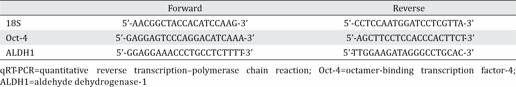
The relative gene expression quantitation of Oct-4 and ALDH1 was measured by using the qRT-PCR-derived cDNA copy number amplification product. Relative expression was measured using Livak method with 2-ΔΔCT equation.
RESULTS
Relative expression of Oct-4 mRNA
Figure 1 shows that Oct-4 mRNA expression in keloid tissues was lower by a half compared with that in normal preputial skins.
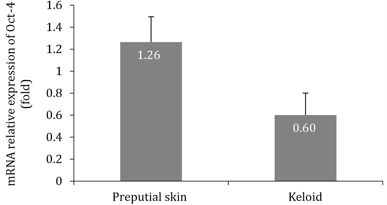
Figure 1. Relative expressions of Oct-4 mRNA in preputial skin and keloid tissue showed insignificant difference (p=0.485; t-test).
Relative expression of ALDH1 mRNA
Figure 2 shows that ALDH1 mRNA expression in keloid tissues was lower by threefourths compared with that in normal preputial skins.
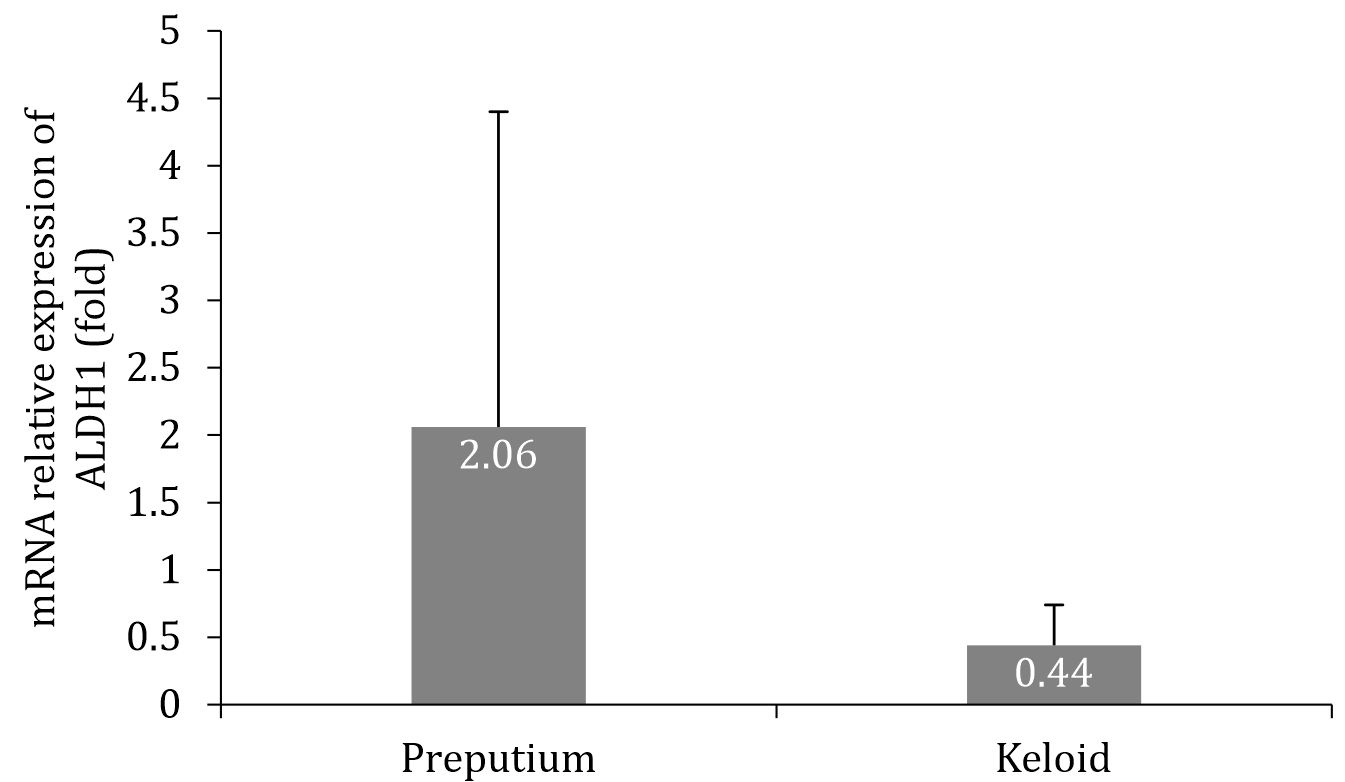
Figure 2. Relative expression of ALDH1 mRNA in preputial skin and keloid tissue showed insignificant difference (p=0.127; t-test).
DISCUSSION
Oct-4, one of the widely used stemness markers, is a master pluripotency and embryonic stem cell regulating gene. A previous study proved that high expression of Oct-4 is consistently associated with cancer stem cell (CSC)-like populations responsible for recurrent and resistant diseases.12 Oct-4 was positively identified from human keloid-derived MSCs6 and microvessels of keloid-associated lymphoid tissue (KALT).7
Similar to Oct-4, Nakahata et al stated ALDH1 as a marker for CSC and was identified in different types of pediatric solid tumors.13 Up-regulation of ALDH1 was identified in keloid scar; this result possibly indicated the presence of CSC-like population within the scar.14 ALDH1 was also positively identified in keloid keratinocytes15 and keloid-derived stem cells (KDSC).16 The existence of stem cell markers in keloid may play Table 1. Primer pairs used for qRT-PCR qRT-PCR=quantitative reverse transcription–polymerase chain reaction; Oct-4=octamer-binding transcription factor-4; ALDH1=aldehyde dehydrogenase-1 Figure 1. Relative expressions of Oct-4 mRNA in preputial skin and keloid tissue showed insignificant difference (p=0.485; t-test). an important role in keloid pathogenesis.
This study showed the expressions of Oct- 4 and ALDH1 in keloid and in normal skin. The expressions of Oct-4 and ALDH1 mRNA decreased in keloid tissues compared with those in normal skin. These results suggest the lower stemness of keloid tissue than normal skin. The low expressions of Oct-4 and ALDH1 mRNA in keloid tissue could be due to the excellent differentiation of keloid stromal cells into myofibroblast in comparison with normal skin cells. In the wound healing response, keloid formation is preceded by activation of immune cells and fibroblasts, which act as the major players in keloid growth.17 The differentiation process was identified by upreregulation of collagen-1, alpha-smooth muscle actin (α-SMA), and thrombospondin-1 in keloid fibroblast culture.18 Wulandari et al19 also showed the up-regulation of collagen-1 and collagen-3 at the mRNA and protein levels in keloid tissues.
Based on previous studies,2keloid possesses a similar characteristic with benign tumors. Hence, keloid is assumed to possess higher stemness characteristic compared with normal tissues. By contrast, the results of this study showed that keloid tissue exhibited similar stemness characteristic to normal tissues. This result might be due to the different ages of the control and keloid subjects. The keloid subjects’ mean age was much higher than that of the control subjects (26 vs 5 years old). Therefore, this discrepancy might explain the lower stemness markers in keloid subjects. In addition, the levels of expressions of Oct-4 and ALDH1 in one control subject were extremely high compared with those of other control and keloid subjects. The low expressions of stemness markers in this study might be proposed to predict low keloid recurrence, as Oct-4 and ALDH1 are associated with cancer recurrence.12However, a further cohort study is required to prove the association between stemness markers and keloid recurrence.
This study presented several limitations. First, normal tissues as controls were not derived from the same subjects. Each donor may present different patterns of stemness characteristics due to the differences in age and location of scar. Also, the number of samples was limited as most keloid patients prefer to undergo other therapies such as topical steroid treatment rather than keloid surgery. The other limitation was sample preparation, in which separating the dermis and epidermis using a scissor and tweezer could remove the reticular dermis. On the other hand, other studies stated that KDSC are located in the reticular dermis, which is located directly beneath the epidermis layer.3,7
This study concludes that keloid tissues express Oct-4 and ALDH1 as stemness markers, and the stemness characteristics of keloid might be similar to a normal skin.
Conflicts of Interest
Sri W.A. Jusman is one of the editorial board members, but was not involved in the review or decision process of the article.
Acknowledgment
Researchers would like to express their gratitude to Direktorat Riset dan Pengabdian Masyarakat UI, who funded this research through Hibah Publikasi Ilmiah Terindeks Tugas Akhir (PITTA).
REFERENCES
- Goder M, Kornhaber R, Bordoni D, Winkler E, Haik J, Tessone A. Cutaneous basal cell carcinoma arising within a keloid scar: a case report. Onco Targets Ther. 2016;9:4793–6
- Ma X, Chen J, Xu B, Long X, Qin H, Zhao RC, et al. Keloid-derived keratinocytes acquire a fibroblastlike appearance and an enhanced invasive capacity in a hypoxic microenvironment in vitro. Int J Mol Med. 2015;35(5):1246–56.
- Zhang Q, Yidi W, Ann DK, Messadi DV, Tuan T, Kelly AP, et al. Mechanisms of hypoxic regulation of plasminogen activator inhibitor-1 gene expression in keloid fibroblasts. J Invest Dermatol. 2003;121(5):1005–12.
- Zeineddine D, Hammoud AA, Mortada M, Boeuf H. The Oct4 protein: more than a magic stemness marker. Am J Stem Cells. 2014;3(2):74–82.
- Samardzija C, Michael Q, Jock KF, Nuzhat A. Attributes of Oct4 in stem cell biology: perspectives on cancer stem cells of the ovary. J Ovarian Res. 2012;5(1):37–42.
- Deng C, Wang B, Zhang Z, Sun G, Zhu J, Wang D, et al. The expressions of notch genes in human keloid-derived mesenchymal-like stem cells. Zhonghua Zheng Xing Wai Ke Za Zhi. 2014;30(3):197–202.
- Grant C, Chudakova DA, Itinteang T, Chibnall AM, Brasch HD, Davis PF, et al. Expression of embryonic stem cell markers in keloid-associated lymphoid tissue. J Clin Pathol. 2016;69(7):643–6.
- Douville J, Beaulieu R, Balicki D. ALDH1 as a functional marker of cancer stem and progenitor cells. Stem Cells Dev. 2009;18(1):17–26.
- Lohberger B, Beate R, Nicole S, Markus A, Bernadette LA, Sonja MW, et al. Aldehyde dehydrogenase 1, a potential marker for cancer stem cells in human sarcoma. PLoS One. 2012;7(8):1–10.
- Hahn JM, Glaser K, McFarland KL, Aronow BJ, Boyce ST, Supp DM. Keloid-derived keratinocytes exhibit an abnormal gene expression profile consistent with a distinct causal role in keloid pathology. Wound Repair Regen. 2013;21(4):530–44.
- Battula VL, Evans KW, Hollier BG, Shi Y, Marini FC, Ayyanan A, et al. Epithelial-mesenchymal transitionderived cells exhibit multilineage differentiation potential similar to mesenchymal stem cells. Stem Cells. 2010;28(8):1435–45.
- Samardzija C, Quinn M, Findlay JK, Ahmed N. Attributes of Oct4 in stem cell biology: perspectives on cancer stem cells of the ovary. J Ovarian Res. 2012;5(37):1–12.
- Nakahata K, Shuichiro U, Shimpei N, Miyoko K, Masahiro Z, Takaharu O, et al. Aldehyde dehydrogenase 1 (ALDH1) is a potential marker for cancer stem cells in embryonal rhabdomyosarcoma. PLoS One. 2015;10(4):1–16.
- Jumper N, Hodgkinson T, Paus R, Bayat A. Site-specific gene expression profiling as a novel strategy for unravelling keloid disease pathobiology. PLoS One. 2017;12(3):1–33.
- Yan L, Rui C, Yuan BL, Lian ZW, Bo P, Xiao YL, et al. MiR-21-5p links epithelial-mMesenchymal transition phenotype with stem-like cell signatures via AKT signaling in keloid keratinocytes. Sci Rep. 2016;6:28281.
- Wang DL, Zhu JJ, Deng CL, Wang B, Yu LM. Identification of biological characteristics of human keloid-derived stem cells. Zhonghua Shao Shang Za Zhi. 2011;27(3):210–4.
- Rodemann HP, Rennekampff HO. Functional diversity of fibroblast. In: Mueller MM, Fusenig NE editors. Tumorassociated fibroblast and their matrix. New York: Springer; 2014. P.23-36.
- Chipev, Simman R, Hatch G, Katz AE, Siegel DM, Simon M. Myofibroblast phenotype and apoptosis in keloid and palmar fibroblasts invitro. Cell Death Differ. 2000;7:166–76.
- Wulandari E, Jusman SW, Moenadjat Y, Jusuf AA, Sadikin M. Expressions of collagen I and III in hypoxic keloid tissue. Kobe J Med Sci. 2016;62(3):E58–69.
Copyright @ 2018 Authors. This is an open access article distributed under the terms of the Creative Commons Attribution-NonCommercial 4.0 International License (http://creativecommons.org/licenses/by-nc/4.0/), which permits unrestricted non-commercial use, distribution, and reproduction in any medium, provided the original author and source are properly cited.
mji.ui.ac.id