Isolated petrous apex ectopic craniopharyngioma
DOI:
https://doi.org/10.13181/mji.v24i4.1261Keywords:
petrous apex tumor, primary ectopic craniopharyngioma, transphenoidAbstract
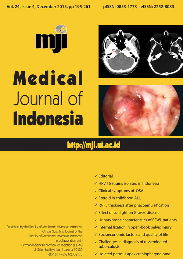
Primary ectopic craniopharyngioma is a rare entity. Isolated petrous apex bone location has not been reported previously. This study reports a case of 26-year-old male with right abducent nerve palsy. CT and MRI imaging reveal right petrous apex cystic lesion. No sellar or suprasellar region involvement was found. Endoscopic endonasal transphenoid approach has been successfully performed. Histopathology examination confirms the diagnosis of adamantinomatous craniopharyngioma. So far, itâs probably the first case report of primary ectopic craniopharyngioma isolated in the petrous apex. This case report supports the premise that primary ectopic craniopharyngioma is a multifactorial process that starts with an error from migrated embryological cells.Downloads
References
Yu X, Liu R, Wang Y, Wang H, Zhao H, Wu Z. Infrasellar craniopharyngioma. Clin Neurol Neurosurg. 2012;114(2):112–9. http://dx.doi.org/10.1016/j.clineuro.2011.09.010
Khalatbari MR, Borghei-Razavi H, Samadian M, Moharamzad Y, Schick U. Isolated primary craniopharyngioma in the cerebellopontine angle. J Clin Neurosci. 2012;19(11):1516–9. http://dx.doi.org/10.1016/j.jocn.2011.09.029
Jakobs M, Orakcioglu B. Ectopic recurrent craniopharyngioma of the frontal bone. J Neurosurg. 2012;117(3):490–7. http://dx.doi.org/10.3171/2012.6.JNS111885
Senthilvel HN, Krishnan SS, Vasudevan MC. Extracranial infrasellar craniopharyngioma. Neurol India. 2014;62(1): 100–3. http://dx.doi.org/10.4103/0028-3886.128356
Banczerowski P, Bálint K, Sipos L. Temporal extradural ectopic craniopharyngioma. Case report. J Neurosurg. 2007;107(1):178–80. http://dx.doi.org/10.3171/JNS-07/07/0178
Yan Y, Tang WY, Yang G, Zhong D. Isolated cerebellopontine angle craniopharyngioma. J Clin Neurosci. 2009;16(12):1655–7. http://dx.doi.org/10.1016/j.jocn.2009.02.029
Ortega-Porcayo LA, Ponce-Gómez JA, Martínez-Moreno M, Portocarrero-Ortíz L, Tena-Suck ML, Gómez-Amador JL. Primary ectopic frontotemporal craniopharyngioma. Int J Surg Case Rep. 2015;9:57–60. http://dx.doi.org/10.1016/j.ijscr.2014.12.038
Bianco Ade M, Madeira LV, Rosemberg S, Shibata MK. Cortical seeding of a craniopharyngioma after craniotomy: case report. Surg Neurol. 2006;66(4):437–40. http://dx.doi.org/10.1016/j.surneu.2005.12.027
Bozbuga M1, Turan Suslu H, Hicdonmez T, Bayindir C. Primary cerebellopontine angle craniopharyngioma in a patient with Gardner syndrome. J Clin Neurosci. 2011;18(2):300–1. http://dx.doi.org/10.1016/j.jocn.2010.06.006
Kawamata T, Kubo O, Kamikawa S, Hori T. Ectopic clival craniopharyngioma. Acta Neurochir (Wien). 2002;144(11):1221–4. http://dx.doi.org/10.1007/s00701-002-1022-6
Lee YH, Kim SD, Lim DJ, Park JY, Chung YG, Kim YS. Isolated petroclival craniopharyngioma with aggressive skull base destruction. Yonsei Med J. 2009;50(5):729–31. http://dx.doi.org/10.3349/ymj.2009.50.5.729
Zada G, Lin N, Ojerholm E, Ramkissoon S, Laws ER. Craniopharyngioma and other cystic epithelial lesions of the sellar region: a review of clinical, imaging, and histopathological relationships. Neurosurg Focus. 2010;28(4): E4. http://dx.doi.org/10.3171/2010.2.FOCUS09318
Downloads
Published
How to Cite
Issue
Section
License
Authors who publish with Medical Journal of Indonesia agree to the following terms:
- Authors retain copyright and grant Medical Journal of Indonesia right of first publication with the work simultaneously licensed under a Creative Commons Attribution-NonCommercial License that allows others to remix, adapt, build upon the work non-commercially with an acknowledgment of the work’s authorship and initial publication in Medical Journal of Indonesia.
- Authors are permitted to copy and redistribute the journal's published version of the work non-commercially (e.g., post it to an institutional repository or publish it in a book), with an acknowledgment of its initial publication in Medical Journal of Indonesia.


