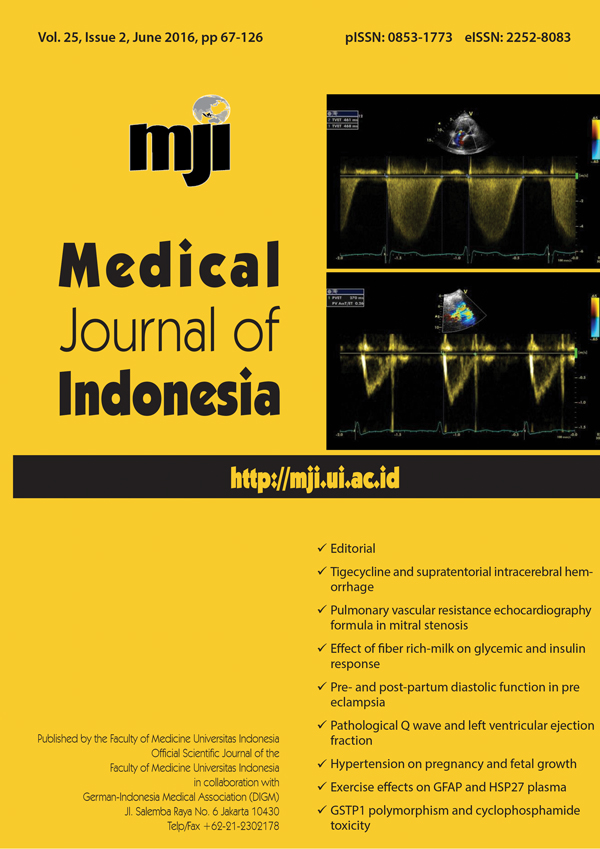
Diastolic function in patients with preeclampsia during pre- and post-partum period using tissue doppler imaging
DOI:
https://doi.org/10.13181/mji.v25i2.1410Keywords:
diastolic dysfunction, postpartum, preeclampsia, prepartumAbstract
Background: Prior studies have compared the E/e’ elevation in preeclampsia patients to normal patients, however there are no data whether this elevation persists after birth. The aim of this study is to analyze diastolic function in preeclampsia patients during pre- and post-partum period using E/e’ parameter measurement.
Methods: This is a prospective cohort study of pregnant women with preeclampsia who were hospitalized and planned for pregnancy termination. Basic clinical characteristics were obtained from all samples. Echocardiography was done prepartum, 48-72 hours after termination, and 40-60 days postpartum. Post hoc analysis using least significant difference method was used to compare the results between measurements.
Results: 30 subjects were enrolled in the study. Analysis on E/e’ characteristics showed statistical difference between prepartum E/e’ and 40 days postpartum E/e’ (11.87±3.184 vs 9.43±2.529, p=0.001, CI=1.123-3.751), as well as between 48 hours post-partum and 40 days post-partum period (12.12±2.754 vs 9.43±2.529, p<0.001, CI=1.615-3.771). There were no statistical differences between pre-partum E/e’ and 48 hours post-partum E/e’ (11.87±3.184 vs 12.12±2.754, p=0.633, CI=-1.345-0.832).
Conclusion: This study showed diastolic dysfunction in preeclampsia patients persists up until a few days after birth, but resolves in time (40 days after birth) as measured by tissue doppler imaging.
Downloads
References
Naidoo DP, Fayers S, Moodley J. Cardiovascular haemodynamics in pre-eclampsia using brain naturetic peptide and tissue Doppler studies. Cardiovasc J Afr. 2013;24(4):130-6. http://dx.doi.org/10.5830/CVJA-2013-023
Sibai B, Dekker G, Kupferminc M. Pre-eclampsia. Lancet. 2005;365(9461):785-99. http://dx.doi.org/10.1016/S0140-6736(05)71003-5
Regitz-Zagrosek V, Lundqvist CB, Borghi C, Cifkova R, Ferreira R, Foidart JM, et al. ESC guidelines on the management of cardiovascular diseases during pregnancy: the task force on the management of cardiovascular diseases during pregnancy of the European Society of Cardiology (ESC). Eur Heart J. 2011;32:3147-97. http://dx.doi.org/10.1093/eurheartj/ehr218
Mor-Avi V, Lang RM, Badano LP, Belohlavek M, Cardim NM, Derumeaux, et al. Current and evolving echocardiographic techniques for the quantitative evaluation of cardiac mechanics: ASE/EAE consensus statement on methodology and indications endorsed by the Japanese Society of Echocardiography. J Am Soc Echocardiogr. 2011;24(3):277-313. http://dx.doi.org/10.1016/j.echo.2011.01.015
Nagueh SF, Appleton CP, Gillebert TC, Marino PN, Oh JK, Smiseth OA, et al. Recommendations for the evaluation of left ventricular diastolic function by echocardiography. J Am Soc Echocardiogr. 2009;22(2):107-33. http://dx.doi.org/10.1016/j.echo.2008.11.023
Kasner M, Gaub R, Sinning D, Westermann D, Steendijk P, Hoffmann W, et al. Global strain rate imaging for the estimation of diastolic function in HFNEF compared with pressure-volume loop analysis. Eur J Echocardiogr. 2010;11(9):743-51. http://dx.doi.org/10.1093/ejechocard/jeq060
Kossaify A, Nicolas N. Impact of overweight and obesity on left ventricular diastolic function and value of tissue doppler echocardiography. Clin Med Insights Cardiol. 2013;7:43-50. http://dx.doi.org/10.4137/CMC.S11156
Firstenberg MS, Levine BD, Garcia MJ, Greenberg NL, Cardon L, Morehead AJ, et al. Relationship of echocardiographic indices to pulmonary capillary wedge pressures in healthy volunteers. J Am Coll Cardiol. 2000;36(5):1664-9. http://dx.doi.org/10.1016/S0735-1097(00)00909-8
Burns AT, Connelly KA, La Gerche A, Mooney DJ, Chan J, Maclsaac AI, et al. Effect of heart rate on tissue Doppler measures of diastolic function. Echocardiography. 2007;24(7):697-701. http://dx.doi.org/10.1111/j.1540-8175.2007.00466.x
Ando T, Kaur R, Holmes AA, Brusati A, Fujikura K, Taub CC. Physiological adaptation of the left ventricle during the second and third trimesters of a healthy pregnancy: a speckle tracking echocardiography study. Am J Cardiovasc Dis. 2015;5(2):119-26.
Rafik HR, Larsson A, Pernow J, Bremme K, Eriksson MJ. Assessment of left ventricular structure and function in preeclampsia by echocardiography and cardiovascular biomarkers. J Hypertens. 2009;27(11):2257-64. http://dx.doi.org/10.1097/HJH.0b013e3283300541
Mammaro A, Carrara S, Cavaliere A, Ermito S, Dinatale A, Pappalardo EM, et al. Hypertensive disorders of pregnancy. J Prenat Med. 2009;3(1):1-5.
Ghossein-Doha C, Peeters L, van Heijster S, van Kuijk S, Spaan J, Delhaas T, et al. Hypertension after preeclampsia is preceded by changes in cardiac structure and function. Hypertension. 2013;62(2):382-90. http://dx.doi.org/10.1161/HYPERTENSIONAHA.113.01319
San-Frutos L, Engels V, Zapardiel I, Perez-Medina T, Almagro-Martinez J, Fernandez R, et al. Hemodynamic changes during pregnancy and postpartum: a prospective study using thoracic electrical bioimpedance. J Matern Fetal Neonatal Med. 2011;24(11):1333-40. http://dx.doi.org/10.3109/14767058.2011.556203
Published
How to Cite
Issue
Section
License
Authors who publish with Medical Journal of Indonesia agree to the following terms:
- Authors retain copyright and grant Medical Journal of Indonesia right of first publication with the work simultaneously licensed under a Creative Commons Attribution-NonCommercial License that allows others to remix, adapt, build upon the work non-commercially with an acknowledgment of the work’s authorship and initial publication in Medical Journal of Indonesia.
- Authors are permitted to copy and redistribute the journal's published version of the work non-commercially (e.g., post it to an institutional repository or publish it in a book), with an acknowledgment of its initial publication in Medical Journal of Indonesia.


