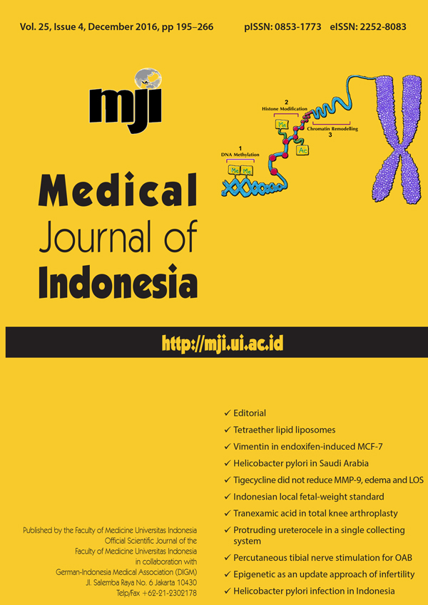
Epigenetic: A new approach to etiology of infertility
DOI:
https://doi.org/10.13181/mji.v25i4.1504Keywords:
epigenetic alteration, epigenetic modification, female infertility, male infertilityAbstract
Infertility is a complex disease which could be caused by male and female factors. The etiology from both factors needs further study. There are some approaches to understanding the etiology of infertility, one of them is epigenetic. Epigenetic modifications consist of DNA methylation, histone modifications, and chromatin remodelling. Male and female germinal cells undergo epigenetic modifications dynamically during differentiation into matured sperm and oocyte cells. In a male, the alteration of DNA methylation in spermatogenesis will cause oligo/asthenozoospermia. In addition, the histone methylation, acetylation, or other histone modification may lead sperm lose its ability to fertilize oocyte. Similarly, in a female, the alteration of DNA methylation and histone modification affects oogenesis, created aneuploidy in fertilized oocytes and resulted in embryonic death in the uterus. Alteration of these epigenetic modification patterns will cause infertility, both in male and female.Downloads
References
Stanford JB, Parnell TA, Boyle PC. Outcomes from treatment of infertility with natural procreative technology in an Irish general practice. J Am Board Fam Med. 2008;21(5):375–84. https://doi.org/10.3122/jabfm.2008.05.070239
Carrell DT. Epigenetics of the male gamete. Fertil Steril. 2012;97(2):267–74. https://doi.org/10.1016/j.fertnstert.2011.12.036
Güne? S, Kulaç T. The role of epigenetics in spermatogenesis. Turk J Urol. 2013;39(3):181–7. https://doi.org/10.5152/tud.2013.037
Zamudio NM, Chong S, O'Bryan MK. Epigenetic regulation in male germ cells. Reproduction. 2008;136(2):131–46. https://doi.org/10.1530/REP-07-0576
Armstrong L. Epigenetics. New York: Garland Science; 2014.
Tollefsbol TO. Handbook of epigenetics: the new molecular and medical genetics. In: Tollefsbol TO, editor. Epigenetics: the new science of genetics. London: Academic Press; 2010. p. 1–6.
Rose NR, Klose RJ. Understanding the relationship between DNA methylation and histone lysine methylation. Biochim Biophys Acta. 2014;1839(12):1362–72. https://doi.org/10.1016/j.bbagrm.2014.02.007
Deaton AM, Bird A. CpG islands and the regulation of transcription. Genes & Dev. 2011;25(10):1010–22. https://doi.org/10.1101/gad.2037511
Okano M, Xie S, Li E. Cloning and characterization of a family of novel mammalian DNA (cytosine-5) methyltransferases. Nat Genet. 1998;19(3):219–20. https://doi.org/10.1038/890
Oh JH, Gertych A, Tajbakhsh J. Nuclear DNA methylation and chromatin condensation phenotypes are distinct between normally proliferating/aging, rapidly growing/immortal, and senescent cells. Oncotarget. 2013;4(3):474–93. https://doi.org/10.18632/oncotarget.942
Rajender S, Avery K, Agarwal A. Epigenetics, spermatogenesis and male infertility. Mutat Res. 2011;727(3):62–71. https://doi.org/10.1016/j.mrrev.2011.04.002
Stein AB, Jones TA, Herron TJ, Patel SR, Day SM, Noujaim SF, et al. Loss of H3K4 methylation destabilizes gene expression patterns and physiological functions in adult murine cardiomyocytes. J Clin Invest. 2011;121(7):2641–50. https://doi.org/10.1172/JCI44641
Peserico A, Simone C. Physical and functional HAT/HDAC interplay regulates protein acetylation balance. J Biomed Biotechnol. 2011(371832):1–10 https://doi.org/10.1155/2011/371832
Unnikrishnan A, Gafken PR, Tsukiyama T. Dynamic changes in histone acetylation regulate origins of DNA replication. Nat Struct Mol Biol. 2010;17(4):430–7 https://doi.org/10.1038/nsmb.1780
Berger SL. Histone modifications in transcriptional regulation. Curr Opin Genet Dev. 2002;12(2):142–8. https://doi.org/10.1016/S0959-437X(02)00279-4
Shiio Y, Eisenman RN. Histone sumoylation is associated with transcriptional repression. Proc Natl Acad Sci U S A. 2003;100(23):13225–30. https://doi.org/10.1073/pnas.1735528100
Narlikar GJ, Sundaramoorthy R, Owen-Hughes T. Mechanisms and functions of ATP-dependent chromatin-remodeling enzymes. Cell. 2013;154(3):490–503. https://doi.org/10.1016/j.cell.2013.07.011
Tsukiyama T. The in vivo functions of ATP-dependent chromatin-remodelling factors. Nat Rev Mol Cell Bio. 2002;3(6):422–9. https://doi.org/10.1038/nrm828
Lusser A, Kadonaga JT. Chromatin remodeling by ATP-dependent molecular machines. Bioessays. 2003;25(12):1192â??200. https://doi.org/10.1002/bies.10359
La Salle S, Trasler JM. Dynamic expression of DNMT3a and DNMT3b isoforms during male germ cell development in the mouse. Dev Biol. 2006;296(1):71–82. https://doi.org/10.1016/j.ydbio.2006.04.436
Schütte B, El Hajj N, Kuhtz J, Nanda I, Gromoll J, Hahn T, et al. Broad DNA methylation changes of spermatogenesis, inflammation and immune response-related genes in a subgroup of sperm samples for assisted reproduction. Andrology. 2013;1(6):822–9. https://doi.org/10.1111/j.2047-2927.2013.00122.x
Montjean D, Zini A, Ravel C, Belloc S, Dalleac A, Copin H, et al. Sperm global DNA methylation level: association with semen parameters and genome integrity. Andrology. 2015;3(2):235–40. https://doi.org/10.1111/andr.12001
Godmann M, Auger V, Ferraroni-Aguiar V, Di Sauro A, Sette C, Behr R, et al. Dynamic regulation of histone H3 methylation at lysine 4 in mammalian spermatogenesis. Biol Reprod. 2007;77(5):754–64. https://doi.org/10.1095/biolreprod.107.062265
Tachibana M, Nozaki M, Takeda N, Shinkai Y. Functional dynamics of H3K9 methylation during meiotic prophase progression. EMBO J. 2007;26(14):3346–59. https://doi.org/10.1038/sj.emboj.7601767
Okada Y, Tateishi K, Zhang Y. Histone demethylase JHDM2A is involved in male infertility and obesity. J Androl. 2010;31(1):75–8. https://doi.org/10.2164/jandrol.109.008052
Sonnack V, Failing K, Bergmann M, Steger K. Expression of hyperacetylated histone H4 during normal and impaired human spermatogenesis. Andrologia. 2002;34(6):384–90. https://doi.org/10.1046/j.1439-0272.2002.00524.x
Sin HS, Barski A, Zhang F, Kartashov AV, Nussenzweig A, Chen J, et al. RNF8 regulates active epigenetic modifications and escape gene activation from inactive sex chromosomes in post-meiotic spermatids. Genes Dev. 2012;26(24):2737–48. https://doi.org/10.1101/gad.202713.112
Lu LY, Wu J, Ye L, Gavrilina GB, Saunders TL, Yu X. RNF8-dependent histone modifications regulate nucleosome removal during spermatogenesis. Dev Cell. 2010;18(3):371–84. https://doi.org/10.1016/j.devcel.2010.01.010
Xiao Y, Pollack D, Andrusier M, Levy A, Callaway M, Nieves E, et al. Identification of cell-specific targets of sumoylation during mouse spermatogenesis. Reproduction. 2016;151(2):149–66. https://doi.org/10.1530/REP-15-0239
Vigodner M, Morris PL. Testicular expression of small ubiquitin-related modifier-1 (SUMO-1) supports multiple roles in spermatogenesis: silencing of sex chromosomes in spermatocytes, spermatid microtubule nucleation, and nuclear reshaping. Dev Biol. 2005;282(2):480–92. https://doi.org/10.1016/j.ydbio.2005.03.034
Derman S, Seifer DB. Evaluation of female infertility. In: Alvero R, editor. Reproductive endocrinology and infertility. Philadelphia: Mosby; 2007. p. 155–68. https://doi.org/10.1016/B978-0-323-04054-9.50015-0
Simpson J. Molecular approach to common causes of female infertility. Best Pract Res Clin Obstet Gynaecol. 2002;16(5):685–702. https://doi.org/10.1053/beog.2002.0317
Zuccotti M, Merico V, Redi CA, Garagna S. An epigenomic biography of the mammalian oocyte. In: Coticchio G, Albertini DF, Santis DL, editor. Oogenesis. London: Springer; 2013. p. 141–50. https://doi.org/10.1007/978-0-85729-826-3_10
Kageyama S, Liu H, Kaneko N, Ooga M, Nagata M, Aoki F. Alterations in epigenetic modifications during oocyte growth in mice. Reproduction. 2007;133(1):85–94. https://doi.org/10.1530/REP-06-0025
Hiura H, Obata Y, Komiyama J, Shirai M, Kono T. Oocyte growth-dependent progression of maternal imprinting in mice. Genes Cells. 2006;11(4):353–61. https://doi.org/10.1111/j.1365-2443.2006.00943.x
Gu L, Wang Q, Sun Q-Y. Histone modifications during mammalian oocyte maturation: dynamics, regulation and functions. Cell Cycle. 2010;9(10):1942–50. https://doi.org/10.4161/cc.9.10.11599
Pan Z, Zhang J, Li Q, Li Y, Shi F, Xie Z, et al. Current advances in epigenetic modification and alteration during mammalian ovarian folliculogenesis. J Genet Genomics. 2012;39(3):111–23. https://doi.org/10.1016/j.jgg.2012.02.004
Hamatani T, Carter MG, Sharov AA, Ko MS. Dynamics of global gene expression changes during mouse preimplantation development. Dev Cell. 2004;6(1):117–31. https://doi.org/10.1016/S1534-5807(03)00373-3
Yue MX, Fu XW, Zhou GB, Hou YP, DU M, Wang L, et al. Abnormal DNA methylation in oocytes could be associated with a decrease in reproductive potential in old mice. J Assist Reprod Genet. 2012;29(7):643–50. https://doi.org/10.1007/s10815-012-9780-4
Akiyama T, Nagata M, Aoki F. Inadequate histone deacetylation during oocyte meiosis causes aneuploidy and embryo death in mice. Proc Natl Acad Sci U S A. 2006;103(19):7339–44. https://doi.org/10.1073/pnas.0510946103
Pons D, de Vries FR, van den Elsen PJ, Heijmans BT, Quax PH, Jukema JW. Epigenetic histone acetylation modifiers in vascular remodelling: new targets for therapy in cardiovascular disease. Eur Heart J. 2009;30(3):266–77. https://doi.org/10.1093/eurheartj/ehn603
Rossetto D, Avvakumov N, Côté J. Histone phosphorylation: a chromatin modification involved in diverse nuclear events. Epigenetics. 2012;7(10):1098–108. https://doi.org/10.4161/epi.21975
Sawicka A, Seiser C. Sensing core histone phosphorylation - a matter of perfect timing. Biochim Biophys Acta. 2014;1839(8):711–8. https://doi.org/10.1016/j.bbagrm.2014.04.013
Li Z, Cao R, Wang M, Myers MP, Zhang Y, Xu RM. Structure of a Bmi-1-Ring1B polycomb group ubiquitin ligase complex. J Biol Chem. 2006;281(29):20643–9. https://doi.org/10.1074/jbc.M602461200
Naqvi H, Ilagan Y, Krikun G, Taylor HS. Altered genome-wide methylation in endometriosis. Reprod Sci. 2014;21(10):1237–43. https://doi.org/10.1177/1933719114532841
Nasu K, Kawano Y, Tsukamoto Y, Takano M, Takai N, Li H, et al. Aberrant DNA methylation status of endometriosis: epigenetics as the pathogenesis, biomarker and therapeutic target. J Obstet Gynaecol Res. 2011;37(7):683–95. https://doi.org/10.1111/j.1447-0756.2011.01663.x
Zanatta A, Rocha AM, Carvalho FM, Pereira RM, Taylor HS, Motta EL, et al. The role of the Hoxa10/HOXA10 gene in the etiology of endometriosis and its related infertility: a review. J Assist Reprod Genet. 2010;27(12):701–10. https://doi.org/10.1007/s10815-010-9471-y
Monteiro JB, Colón-Diaz , Garcia M, Gutierrez S, Colón M, Seto E, et al. Endometriosis is characterized by a distinct pattern of histone 3 and histone 4 lysine modifications. Reprod Sci. 2014;21(3):305–18. https://doi.org/10.1177/1933719113497267
Ndefo UA, Eaton A, Green MR. Polycystic ovary syndrome: a review of treatment options with a focus on pharmacological approaches. PT. 2013;38(6):336.
Wang P, Zhao H, Li T, Zhang W, Wu K, Li M, et al. Hypomethylation of the LH/choriogonadotropin receptor promoter region is a potential mechanism underlying susceptibility to polycystic ovary syndrome. Endocrinology. 2014;155(4):1445–52. https://doi.org/10.1210/en.2013-1764
Young JM, McNeilly AS. Theca: the forgotten cell of the ovarian follicle. Reproduction. 2010;140(4):489–504. https://doi.org/10.1530/REP-10-0094
Published
How to Cite
Issue
Section
License
Authors who publish with Medical Journal of Indonesia agree to the following terms:
- Authors retain copyright and grant Medical Journal of Indonesia right of first publication with the work simultaneously licensed under a Creative Commons Attribution-NonCommercial License that allows others to remix, adapt, build upon the work non-commercially with an acknowledgment of the work’s authorship and initial publication in Medical Journal of Indonesia.
- Authors are permitted to copy and redistribute the journal's published version of the work non-commercially (e.g., post it to an institutional repository or publish it in a book), with an acknowledgment of its initial publication in Medical Journal of Indonesia.


