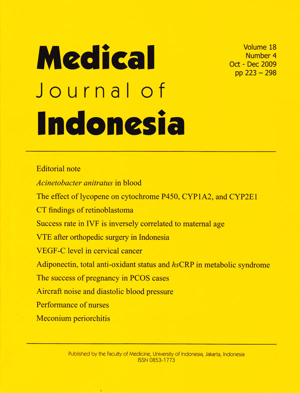
Computed tomography findings of retinoblastoma patients at Cipto Mangunkusumo Hospital Jakarta
DOI:
https://doi.org/10.13181/mji.v18i4.366Keywords:
leukocoria, enhancement, calcificationAbstract
Aim As data of CT findings for retinoblatoma in Indonesia is not yet available, this study aimed to determine patient distribution and to describe CT findings of RB patients at Cipto Mangunkusumo Hospital.
Methods From April 2004 to August 2007, CT scans were performed on 64 new patients of RB with clinical findings: leukocoria (19 patients), leukocoria with proptosis (41 patients), buphthalmus (2 patients) and red eyes (2 patients). The CT can was performed using single slice CT scan and multislice CT scan with axial and coronal section. The cases were categorized into 3 groups: grade I: tumor confined to the globe, grade II: tumor extended to orbital soft tissue or involved the optic nerve, and grade III: tumor extended beyond the orbit or intracranial space. The CT features were evaluated in each group for the presence of calcification, contrast enhancement and lateralization.
Results Sixty four patients (30 females and 34 males) were evaluated. Age range: 3 months up to 9 years old; bilateral 20 patients, unilateral 44 patients. Six patients of 19 patients with clinical finding leukocoria were included in group I, and the remaining were included in group II. Twenty seven patients of 41 patients with proptotic eyes were included in group II, and the remaining were included in group III. Patients with clinical finding buphthalmus and red eyes were included in group II. All of the group I tumors (6 patients) showed calcification and enhancement. In group II (44 patients), calcification was detected in 41 patients, and there was no evidence of contrast enhancement in 6 patients. Group III (14 patients): no calcification in 2 patients, all of the tumors showed enhancement.
Conclusion Our study showed calcification in 92% of RB. Calcification was not a favorable prognostic sign, because calcification was detected in almost all of the extraocular and intracranial tumors. Tumor enhancement was not correlated with extraocular tumor extension. The finding of leukocoria without proptosis could not exclude the presence of extraocular tumor extension. Suspicious intracranial invasion should be considered in proptotic RB patient. Most retinoblastoma cases in Indonesia are diagnosed at advanced stage, so that the objective of the therapy is life saving rather than visual salvation. (Med J Indones 2009; 18: 239-43)
Downloads
References
Gatta G, Capocaccia R, Coleman MP, Ries GA, Berrino F. Childhood cancer survival in Europe and United states. Cancer. 2002; 95(8):1767–72.
Yoo KH, Sohn WY, Sung KW, Jung HL, Koo HH, Oh SY et al. Chemoreduction followed by local therapy and adjuvant chemotherapy for advanced intraocular retinoblastoma. J Korean Med Sci. 2002;17:817–22.
Departemen Anak divisi Hematoonkologi. Data insiden tumor anak. Jakarta:The Department; 2000.
Faerber EN, Poussaint TY. The orbit. In: Kuhn JP, Slovis TL, Haller J, eds. Caffey's pediatric diagnostic imaging. 10th edition. USA: Mosby; 2004. pg. 414-5.
Mafee MF. Eye and orbit. In: Mafee MF, Valvassori GE, Becker M, eds. Imaging of the head and neck. 2nd edition. Germany: Thieme Verlag; 2005. p.137–43.
Barkovich AJ. Intracranial, orbital, and neck masses of childhood. In: Barkovich AJ, ed. Pediatric neuroimaging.
Fourth edition. USA: Lippincott Williams and Wilkins;2005. p.618–20.
Aerts I, Lumbroso le Rovic L, Gauthier Villars M, Brisse H, Doz F, Desjardins L. Retinoblastoma. Orphanet Journal of Rare Diseases. 2006;1:31–40.
Wang SK. Intraocular tumor. In: Wang SK, Lee WR, eds. Ophthalmic pathology. USA:Blackwell; 2005. p.259-60.
Brisse HJ, Lumbroso le Rovic L, Freneanx PC, Validire P, Doz FP, Quintana EJ, et al. CT and MRI findings in diffuse infiltrative retinoblastoma. AJNR. 2001; 22: 499–504.
Olivecrona H, Agerberg AP, Huaman A. CT Diagnosis of retinoblastoma with histopathology correlations. Eur Radiol. 1994; 4:307–13.
Danziger A, Price HI. CT fi ndings in retinoblastoma. AJR. 1979;133:783–5.
Bellaton E, Bertozzi AI, Behar C, Chastagner P, Brisse H, Sainte-Rose C, et al. Neoadjuvant chemotherapy for extensive unilateral retinoblastoma. Br J Ophthalmol. 2003;87:327–29.
Barrett T, Brechbiel M, Bernando M, Peter L, Choyke. MRI of tumor angiogenesis. Journal of MRI. 2007;26:235–49.
Downloads
Published
How to Cite
Issue
Section
License
Authors who publish with Medical Journal of Indonesia agree to the following terms:
- Authors retain copyright and grant Medical Journal of Indonesia right of first publication with the work simultaneously licensed under a Creative Commons Attribution-NonCommercial License that allows others to remix, adapt, build upon the work non-commercially with an acknowledgment of the work’s authorship and initial publication in Medical Journal of Indonesia.
- Authors are permitted to copy and redistribute the journal's published version of the work non-commercially (e.g., post it to an institutional repository or publish it in a book), with an acknowledgment of its initial publication in Medical Journal of Indonesia.


