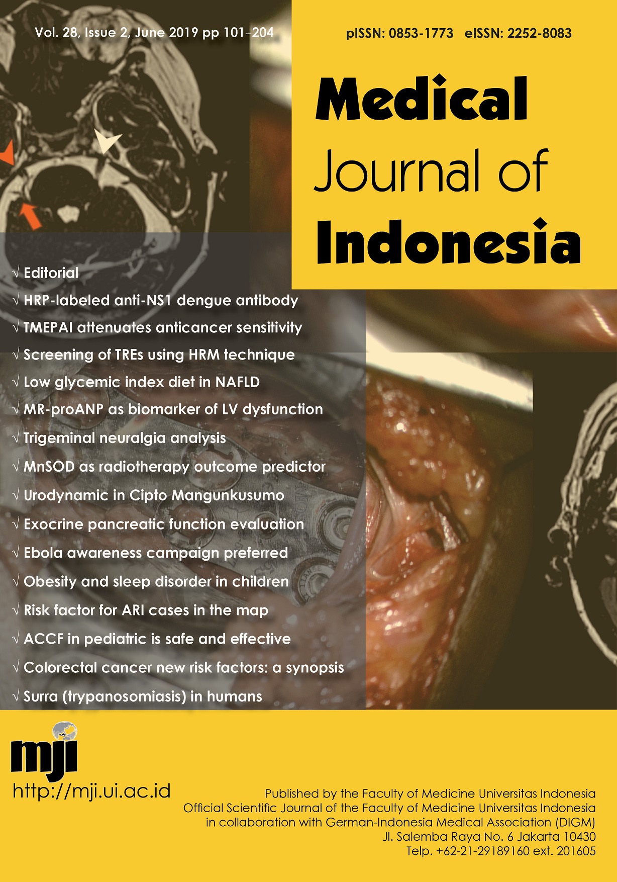
Correlation between pain assessment scale and neurovascular compression distance to the root exit zone in trigeminal neuralgia analysis using 3D CISS MRI sequence
DOI:
https://doi.org/10.13181/mji.v28i2.1796Keywords:
neurovascular compression, pain assessment scale, trigeminal neuralgiaAbstract
BACKGROUND Trigeminal neuralgia (TN) is primarily caused by neurovascular compression (NVC) at root exit zone (REZ) in cerebellopontine angle cistern. In some NVC cases, it was suspected that clinical symptoms may be correlated with the distance of trigeminal nerve root to vascular contact. Pain assessment scale (PAS) was the most common scale used to evaluate TN pain, therefore this study was conducted to analyze the correlation between PAS usingnumeric rating scale (NRS) and distance from the NVC to REZ location in patients with TN using 3D CISS MRI sequence.
METHODS This cross-sectional study was conducted at the Department of Radiology, Faculty of Medicine, Universitas Indonesia, Cipto Mangunkusumo Hospital, Jakarta, using secondary data of 32 patients, collected from Picture Archiving and Communication System from January 2013 to January 2016. Statistical analysis was performed using SPSS, version 20.0. Spearman p-value of < 0.05 was considered significant.
RESULTS A total of 32 patients met the inclusion criteria. The mean (SD) distances from the NVC to the REZ were 2.1 (2.1), 2.31 (2.25), and 3.22 (2.63) mm on the shortest, medial, and lateral sides, respectively. The correlation coefficients (r) between the PAS value and the NVC distance in relation to the trigeminal nerve REZ were -0.39 (p = 0.021), -0.57 (p < 0.01), and -0.57 (p = 0.294) on the shortest, lateral, and medial sides, respectively.
CONCLUSIONS PAS using the NRS instrument exhibited an inverse correlation to NVC distance to the REZ of the trigeminal nerve. Shorter distance increased the PAS value.
Downloads
References
Besta R, Shankar YU, Kumar A, Rajasekhar E, Prakash SB. MRI 3D CISS-a novel imaging modality in diagnosing trigeminal neuralgia-a review. J Clin Diagn Res. 2016;10(3):ZE01-3.https://doi.org/10.7860/JCDR/2016/14011.7348
Joffroy A, Levivier M, Massager N. Trigeminal neuralgia. Pathophysiology and treatment. Acta Neurol Belg. 2001;101(1):20-5.
National Initiative of Pain Control. Wong-Baker FACES Pain Rating Scale. Natl Initiat Pain Control. 2003;2-3.
Chen GQ, Wang XS, Wang L, Zheng JP. Arterial compression of nerve is the primary cause of trigeminal neuralgia. Neurol Sci. 2014;35(1):61-6. https://doi.org/10.1007/s10072-013-1518-2
Antonini G, Di Pasquale A, Cruccu G, Truini A, Morino S, Saltelli G, et al. Magnetic resonance imaging contribution for diagnosing symptomatic neurovascular contact in classical trigeminal neuralgia: a blinded case-control study and meta-analysis. Pain. 2014;155(8):1464-71. https://doi.org/10.1016/j.pain.2014.04.020
Suzuki M, Yoshino N, Shimada M, Tetsumura A, Matsumura T, Fukayama H, et al. Trigeminal neuralgia: differences in magnetic resonance imaging characteristics of neurovascular compression between symptomatic and asymptomatic nerves. Oral Surg Oral Med Oral Pathol Oral Radiol. 2015;119(1):113-8. https://doi.org/10.1016/j.oooo.2014.09.013
Peker S, Kurtkaya O, Üzün ?, Pamir MN. Microanatomy of the central myelin-peripheral myelin transition zone of the trigeminal nerve. Neurosurgery. 2006;59(2):354-9. https://doi.org/10.1227/01.NEU.0000223501.27220.69
Gonçalves FG, do Amaral LL. Constructive interference in steady state imaging in the central nervous system. Eur Neurol Rev. 2011;6(2):138. https://doi.org/10.17925/ENR.2011.06.02.138
Tarnaris A, Renowden S, Coakham HB. A comparison of magnetic resonance angiography and constructive interference in steady state-three-dimensional Fourier transformation magnetic resonance imaging in patients with hemifacial spasm. Br J Neurosurg. 2007;21(4):375-81. https://doi.org/10.1080/02688690701474564
Jo KW, Kong DS, Hong KS, Lee JA, Park K. Long-term prognostic factors for microvascular decompression for trigeminal neuralgia. J Clin Neurosci. 2013;20(3):440-5. https://doi.org/10.1016/j.jocn.2012.03.037
Koh KK. Effects of estrogen on the vascular wall: vasomotor function and inflammation. Cardiovasc Res. 2002;55(4):714-26. https://doi.org/10.1016/S0008-6363(02)00487-X
Cheng J, Meng J, Liu W, Zhang H, Hui X, Lei D. Nerve atrophy in trigeminal neuralgia due to neurovascular compression and its association with surgical outcomes after microvascular decompression. Acta Neurochir. 2017;159(9):1699-705. https://doi.org/10.1007/s00701-017-3250-9
Krzy?ewski RM, Stachura MK, Stachura AM, Rybus J, Tomaszewski KA, Klimek-Piotrowska W, et al. Variations and morphometric analysis of the proximal segment of the superior cerebellar artery. Neurol Neurochir Pol. 2014;48(4):229-35. https://doi.org/10.1016/j.pjnns.2014.07.006
Blitz AM, Northcutt B, Shin J, Aygun N, Herzka DA, Theodros D, et al. Contrast-enhanced CISS imaging for evaluation of neurovascular compression in trigeminal neuralgia: improved correlation with symptoms and prediction of surgical outcomes. Am J Neuroradiol. 2018;39(9):1724-32. https://doi.org/10.3174/ajnr.A5743
Lorenzoni J, David P, Levivier M. Patterns of neurovascular compression in patients with classic trigeminal neuralgia: a high-resolution MRI-based study. Eur J Radiol. 2012;81(8):1851-7. https://doi.org/10.1016/j.ejrad.2009.09.017
Tomii M, Onoue H, Yasue M, Tokudome S, Abe T. Microscopic measurement of the facial nerve root exit zone from central glial myelin to peripheral Schwann cell myelin. J Neurosurg. 2003;99(1):121-4. https://doi.org/10.3171/jns.2003.99.1.0121
Published
How to Cite
Issue
Section
License
Authors who publish with Medical Journal of Indonesia agree to the following terms:
- Authors retain copyright and grant Medical Journal of Indonesia right of first publication with the work simultaneously licensed under a Creative Commons Attribution-NonCommercial License that allows others to remix, adapt, build upon the work non-commercially with an acknowledgment of the work’s authorship and initial publication in Medical Journal of Indonesia.
- Authors are permitted to copy and redistribute the journal's published version of the work non-commercially (e.g., post it to an institutional repository or publish it in a book), with an acknowledgment of its initial publication in Medical Journal of Indonesia.


