

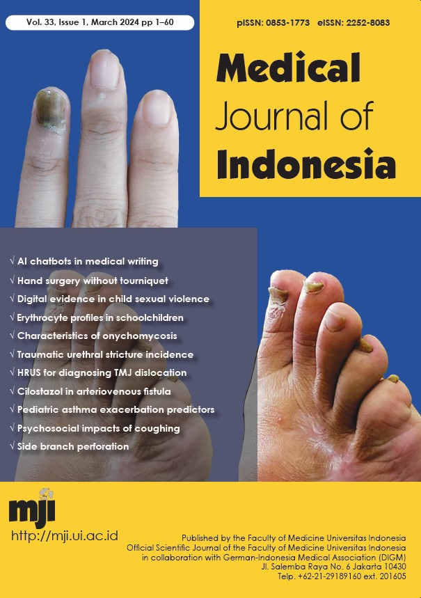
Clinical and microbiological characteristics of onychomycosis in a tertiary hospital: a cross-sectional study
DOI:
https://doi.org/10.13181/mji.oa.247201Keywords:
Candida, microbiology, molds, onychomycosis, prognosis, tertiary hospitalAbstract
BACKGROUND Onychomycosis is a common fungal nail infection with a low cure rate. While dermatophytes are the most common causal agent for onychomycosis, the incidence of Candida and nondermatophyte mold (NDM) onychomycosis is increasing. This study aimed to analyze the clinical and microbiological characteristics of patients with onychomycosis.
METHODS Patients who visited the Department of Dermatology and Venereology, Cipto Mangunkusumo Hospital, and were diagnosed with onychomycosis from 2017 to 2022 were included. Diagnosis was established through clinical examination, supported by the result of direct microscopic examination with potassium hydroxide.
RESULTS Of 171 patients, 93.6% had onychodystrophy, 65.5% were females, and 62.0% were aged 19–59 years. Most patients had onychodystrophy in more than three nails, affecting fingernails (31.6%) and toenails (34.5%). Interestingly, 84.8% of patients had no history of nail diseases. The median onset of disease was 24.0 (1–1,040) weeks, while the median onychomycosis severity index was 10.0 (2–40). Most cases were caused by Candida albicans (48.3%). Fusarium was the only NDM documented (2.3%). Some patients were resistant to itraconazole (11.4%) and miconazole (4.5%). Overall, 49.1% of the patients were declared not cured.
CONCLUSIONS Candida was the predominant cause of onychomycosis, and onychodystrophy was the dominant feature. Current treatment regimens with systemic or topical antifungal agents did not yield satisfactory results, with more than half of the patients deemed not cured.
Downloads
References
Rather S, Keen A, Shah FY, Yaseen A, Farooq S, Bakhshi A. Candidal onychomycosis: clinicoepidemiological profile, prevailing strains, and antifungal susceptibility pattern-a study from a Tertiary Care Hospital. Indian J Dermatol. 2021;66(2):132-7. https://doi.org/10.4103/ijd.IJD_395_20
Fich F, Abarzúa-Araya A, Pérez M, Nauhm Y, León E. Candida parapsilosis and Candida guillermondii: emerging pathogens in nail candidiasis. Indian J Dermatol. 2014;59(1):24-9. https://doi.org/10.4103/0019-5154.123485
Gupta AK, Summerbell RC, Venkataraman M, Quinlan EM. Nondermatophyte mould onychomycosis. J Eur Acad Dermatol Venereol. 2021;35(8):1628-41. https://doi.org/10.1111/jdv.17240
Otaševi? S, Barac A, Pekmezovic M, Tasic S, Ignjatovi? A, Mom?ilovi? S, et al. The prevalence of Candida onychomycosis in Southeastern Serbia from 2011 to 2015. Mycoses. 2016;59(3):167-72. https://doi.org/10.1111/myc.12448
Widaty S, Miranda E, Oktarina C. Candida onychomycosis: mini review. In: Xinhui W, editor. Advances in Candida albicans. Rijeka: IntechOpen; 2021. p. Ch. 5. https://doi.org/10.5772/intechopen.96650
Lipner SR, Scher RK. Onychomycosis: treatment and prevention of recurrence. J Am Acad Dermatol. 2019;80(4):853-67. https://doi.org/10.1016/j.jaad.2018.05.1260
Widaty S, Miranda E, Bramono K, Menaldi SL, Marissa M, Oktarina C, et al. Prognostic factors influencing the treatment outcome of onychomycosis Candida. Mycoses. 2020;63(1):71-7. https://doi.org/10.1111/myc.13018
Karmila ID, Santoso A. Profile of onychomycosis in dermatology outpatient department at Sanglah General Hospital Denpasar, Bali-Indonesia periods 2016-2017. BDV. 2018;1(1):16-9. https://doi.org/10.15562/bdv.v1i1.5
Carney C, Tosti A, Daniel R, Scher R, Rich P, DeCoster J, et al. A new classification system for grading the severity of onychomycosis: onychomycosis severity index. Arch Dermatol. 2011;147(11):1277-82. https://doi.org/10.1001/archdermatol.2011.267
Clinical and Laboratory Standard Institute (CLSI). M54-A: principles and procedures for detection of fungi in clinical specimens-direct examination and culture; approved guideline. Philadelphia: Clinical and Laboratory Standards Institute (CLSI); 2012.
Clinical and Laboratory Standard Institute (CLSI). M61: performance standards for antifungal susceptibility testing of filamentous fungi. Philadelphia: Clinical and Laboratory Standards Institute (CLSI); 2017.
Kabi S, Swain B, Jain S. Epidemiological and microbiological study of onychomycosis. J Clin Diagn Res. 2021;15(3):DC15-8. https://doi.org/10.7860/JCDR/2021/48332.14640
Cho S, Lee H, Hwang JY, Choi JS, Kim HJ, Kim TW, et al. Prevalence and characteristics of onychomycosis in patients with knee osteoarthritis: a single-centre prospective cross-sectional study. Acta Derm Venereol. 2021;101(8):adv00526. https://doi.org/10.2340/00015555-3895
Gupta AK, Versteeg SG, Shear NH. Onychomycosis in the 21st century: an update on diagnosis, epidemiology, and treatment. J Cutan Med Surg. 2017;21(6):525-39. https://doi.org/10.1177/1203475417716362
Cozzani E, Agnoletti AF, Speziari S, Schiavetti I, Zotti M, Persi A, et al. Epidemiological study of onychomycosis in older adults with onychodystrophy. Geriatr Gerontol Int. 2016;16(4):486-91. https://doi.org/10.1111/ggi.12496
Papini M, Piraccini BM, Difonzo E, Brunoro A. Epidemiology of onychomycosis in Italy: prevalence data and risk factor identification. Mycoses. 2015;58(11):659-64. https://doi.org/10.1111/myc.12396
McIntosh IB. Onychodystrophy and onychogryphosis. Podiatry Review. 2021;78:17+.
Sharma R, Saxena R, Sabharwal ER, Mamoria VP. Clinico-mycological profile of onychomycosis: a study from North-Western, India. In: Recent Developments in Medicine and Medical Research. 2021(13):105-12. https://doi.org/10.9734/bpi/rdmmr/v13/15004D
Kayarkatte MN, Singal A, Pandhi D, Das S. Clinico-mycological study of onychomycosis in a tertiary care hospital-a cross-sectional study. Mycoses. 2020;63(1):113-8. https://doi.org/10.1111/myc.13025
Zhang J, Lu S, Huang H, Li X, Cai W, Ma J, et al. Combination therapy for onychomycosis using a fractional 2940-nm Er:YAG laser and 5 % amorolfine lacquer. Lasers Med Sci. 2016;31(7):1391-6. https://doi.org/10.1007/s10103-016-1990-z
El-Tatawy RA, Aliweh HA, Hegab DS, Talaat RAZ, Shams Eldeen MA. Fractional carbon dioxide laser and topical tioconazole in the treatment of fingernail onychomycosis. Lasers Med Sci. 2019;34(9):1873-80. https://doi.org/10.1007/s10103-019-02789-2
Hees H, Jäger MW, Raulin C. Treatment of onychomycosis using the 1 064 nm Nd:YAG laser: a clinical pilot study. J Dtsch Dermatol Ges. 2014;12(4):322-9. https://doi.org/10.1111/ddg.12292
Gupta AK, Venkataraman M, Renaud HJ, Summerbell R, Shear NH, Piguet V. A paradigm shift in the treatment and management of onychomycosis. Skin Appendage Disord. 2021;7(5):351-8. https://doi.org/10.1159/000516112
Gupta AK, Renaud HJ, Quinlan EM, Shear NH, Piguet V. The growing problem of antifungal resistance in onychomycosis and other superficial mycoses. Am J Clin Dermatol. 2021;22(2):149-57. https://doi.org/10.1007/s40257-020-00580-6
Bunyaratavej S, Srinonprasert V, Kiratiwongwan R, Wongdama S, Leeyaphan C. Onychomycosis in older adults: the age and associated factors affecting the complete cure rate. Australas J Dermatol. 2022;63(1):74-80. https://doi.org/10.1111/ajd.13686
Guarner J, Brandt ME. Histopathologic diagnosis of fungal infections in the 21st century. Clin Microbiol Rev. 2011;24(2):247-80. https://doi.org/10.1128/CMR.00053-10
Guilhermetti E, Takahachi G, Shinobu CS, Svidzinski TI. Fusarium spp. as agents of onychomycosis in immunocompetent hosts. Int J Dermatol. 2007;46(8):822-6. https://doi.org/10.1111/j.1365-4632.2007.03120.x
Piraccini BM. Nail disorders due to environmental, professional, and cosmetic causes and auto-induced nail diseases. In: Piraccini BM, editor. Nail disorders: a practical guide to diagnosis and management. Milano: Springer Milan; 2014. p. 55-74. https://doi.org/10.1007/978-88-470-5304-5_6
Downloads
Published
How to Cite
Issue
Section
License
Authors who publish with Medical Journal of Indonesia agree to the following terms:
- Authors retain copyright and grant Medical Journal of Indonesia right of first publication with the work simultaneously licensed under a Creative Commons Attribution-NonCommercial License that allows others to remix, adapt, build upon the work non-commercially with an acknowledgment of the work’s authorship and initial publication in Medical Journal of Indonesia.
- Authors are permitted to copy and redistribute the journal's published version of the work non-commercially (e.g., post it to an institutional repository or publish it in a book), with an acknowledgment of its initial publication in Medical Journal of Indonesia.


