Central nervous system lymphoma: a description and analysis of patients’ clinical and radiological features
Abstract
Background: Central nervous system (CNS) lymphoma is a rare brain neoplasm. Its incidence has increased these years, so it should be considered in the differential diagnosis for mass lesions in the CNS. The aim of the study was to describe the radiological and clinical features of patients with CNS lymphoma.
Methods: The study was a retrospective study. All patients histopathologically confirmed to have CNS lymphoma from November 2008 to December 2013 in Siloam Hospital Lippo Village were included in the study. Medical records and patients’ MRI results were retrieved to be analyzed.
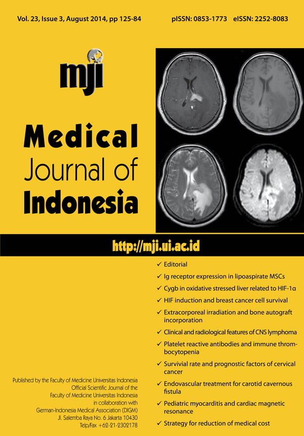
Results: 32 patients were histopathologically diagnosed to have CNS lymphoma. The patients, mean age was 54 ± 15.01 years with slight male predominance. No patient was immuno compromised (CD4 > 500 cells/µL and leukocyte 5,000-11,000 cells/µL). The median interval between the onset of the initial symptoms and diagnosis is 7 weeks. The most common presenting symptoms were headache, mental changes, and neurological deficits related to the location of lesion. MR images show that most lesions were enhanced with contrast, iso-hypointense in T1 weighted imaging (T1WI), iso- to hypointense with perifocal edema in T2 weighted imaging (T2WI), hyperintense in diffusion weighted imaging (DWI), with the most common location was white matter of cerebral hemisphere on one or more lobes and periventricular area, and the tumor could be single or multiple (24%) without clear edges.
Conclusion: Short course of neurological worsening (within weeks) should lead a suspicion toward lymphoma. The characteristics of MR images are markedly enhanced by contrast, iso- to hypointense on T1WI and T2WI, and hyperintense in DWI, involving white matter of cerebral hemisphere and periventricular area.
References
Haldorsen IS, Espeland A, Larsen JL, Mella O. Diagnostic delay in primary central nervous system lymphoma. Acta Oncol. 2005;44(7):728-34. http://dx.doi.org/10.1080/02841860500256272
Morris PG, Abrey LE. Therapeutic challenges in primary CNS lymphoma. Lancet Neurol. 2009;8(6):581-92. http://dx.doi.org/10.1016/S1474-4422(09)70091-2
Olson JE, Janney CA, Rao RD, Cerhan JR, Kurtin PJ, Schiff D, et al. The continuing increase in the incidence of primary central nervous system non-hodgkin lymphoma. Cancer. 2002;95(7):1504-10. http://dx.doi.org/10.1002/cncr.10851
Lister A, Abrey LE, Sandlund JT. Central nervous system lymphoma. ASH Education Book. 2002;2002(1):283-96.
Schabet M. Epidemiology of primary CNS lymphoma. J neurooncol. 1999;43(3):199-201. http://dx.doi.org/10.1023/A:1006290032052
Greenberg MS. Handbook of Neurosurgery. 7th ed. New York: Thieme; 2010. p.1338.
Moradi A, Tajedini A, Mehrabian A, Sadeghi S, Semnani V, Khodabakhshi R, et al. Clinicopathological features of primary central nervous system lymphoma. Neurosciences. 2006;11(4):284-8.
Slone HW, Blake JJ, Shah R, Guttikonda S, Bourekas EC. CT and MRI findings of intracranial lymphoma. AJR Am J Roentgenol. 2005;184(5):1679-85. http://dx.doi.org/10.2214/ajr.184.5.01841679
Haldorsen IS, Espeland A, Larsson EM. Central nervous system lymphoma: characteristic findings on traditional and advanced imaging. AJNR Am J Neuroradiol. 2011;32(6):984-92. http://dx.doi.org/10.3174/ajnr.A2171
Herrlinger U, Schabet M, Bitzer M, Petersen D, Krauseneck P. Primary central nervous system lymphoma: from clinical presentation to diagnosis. J Neurooncol. 1999;43(3):219-26. http://dx.doi.org/10.1023/A:1006298201101
Stadnik TW, Demaerel P, Luypaert RR, Chaskis C, van Rompaey KL, Michotte A, et al. Imaging tutorial: differential diagnosis of bright lesions on diffusion-weighted MR images. Radiographics. 2003;23(1):e7. http://dx.doi.org/10.1148/rg.e7
Toh C-H, Castillo M, Wong AM, Wei KC, Wong HF, Ng SH, et al. Primary cerebral lymphoma and glioblastoma multiforme: differences in diffusion characteristics evaluated with diffusion tensor imaging. AJNR Am J Neuroradiol. 2008;29(3):471-5. http://dx.doi.org/10.3174/ajnr.A0872
Kyritsis AP, Yung WK, Leeds NE, Burner J, Gleason MJ, Levin VA. Multifocal cerebral gliomas associated with secondary malignancies. Lancet. 1992;339(8803):1229-30. http://dx.doi.org/10.1016/0140-6736(92)91167-7
Copyright (c) 2014 Julius July, Andy Wijaya, Priscilla Muliantara, Mira Yuniarti

This work is licensed under a Creative Commons Attribution-NonCommercial 4.0 International License.
Authors who publish with Medical Journal of Indonesia agree to the following terms:
- Authors retain copyright and grant Medical Journal of Indonesia right of first publication with the work simultaneously licensed under a Creative Commons Attribution-NonCommercial License that allows others to remix, adapt, build upon the work non-commercially with an acknowledgment of the work’s authorship and initial publication in Medical Journal of Indonesia.
- Authors are permitted to copy and redistribute the journal's published version of the work non-commercially (e.g., post it to an institutional repository or publish it in a book), with an acknowledgment of its initial publication in Medical Journal of Indonesia.